

Longissimus
By
Della Barnes, an MS Anatomy graduate, blends medical research with accessible writing, simplifying complex anatomy for a better understanding and appreciation of human anatomy.
Last updated:
14/10/2024Della Barnes, MS Anatomy
UX/UI Designer at - AdobeDella Barnes, an MS Anatomy graduate, blends medical research with accessible writing, simplifying complex anatomy for a better understanding and appreciation of human anatomy.
What is the Longissimus Muscle
The longissimus is a deep intrinsic muscle of the back that runs along the sides of the spine, extending from the neck to the lower back. It plays a crucial role in spine and head movements. In Latin, the muscle’s name means “the longest one,” as it is the thickest and longest of the three erector spinae muscles. The other two muscles in the group are the spinalis and iliocostalis.
Anatomy
Location and Attachments
The muscle can be divided into 3 parts based on their points of attachment.
| Origin | Longissimus Capitis: Transverse processes of C4 to T5 vertebrae Longissimus Cervicis: Transverse processes of T1 to T5 vertebrae Longissimus Thoracis: Transverse processes of L1 to L5, and the ilium and sacrum |
| Insertion | Capitis: The temporal bone’s mastoid process Cervicis: Transverse processes of C2 to C6 vertebrae Thoracis: Transverse processes of all the thoracic and lumbar vertebrae |
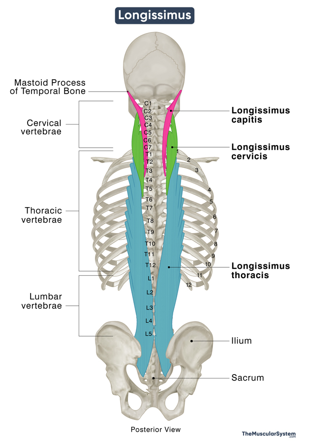
1. Capitis Part
It is the topmost part of the muscle, located at the base of the skull and neck. It originates from the transverse processes of the 5 upper thoracic vertebrae (T1-T5) and the articular processes of the 4 lower cervical vertebrae (C4-C7).
The muscle fibers then travel superiorly and slightly laterally, crossing the semispinalis capitis muscle. They attach to the mastoid process of the temporal bone located directly behind the ear. Additionally, a few fibers of the capitis part also attach to the transverse processes of the lower cervical vertebrae (C4-C7).
2. Cervicis Part
The cervicis part lies in the lower neck and upper back region. Like the capitis part, it also arises from the transverse processes of the first 5 thoracic vertebrae (T1-T5). It then runs upward, between the tendons of the capitis and thoracic parts, to insert into the transverse processes (posterior tubercles) of the 2nd to 6th cervical vertebrae (C2-C6).
3. Thoracis Part
The lowest portion of the longissimus is the thoracis part, which attaches to the vertebrae in the middle to lower back (L1-L5 and pelvic bones). It is the most prominent part of the muscle and is further divided into the thoracic and lumbar regions.
The thoracic region consists of 11-12 fascicles originating from the spinous and transverse processes of all the lumbar vertebrae, the posterior aspect of the sacrum bone, the median sacral crest, and the iliac crest. Its fibers run upwards, extending into rounded tendons that insert into the transverse processes of all the 12 thoracic vertebrae. A few tendons also attach to the 4th or 5th to the 12 ribs, between the angle and tubercle of each rib.
The lumbar region consists of 5 fascicles that originate from the lumbar intermuscular aponeurosis, the sacropelvic surface of the ilium, and the posterior sacroiliac ligament. The fibers are then inserted into the accessory processes on the posterior surface of the transverse processes of all five lumbar vertebrae.
Relations With Surrounding Muscles and Structures
As mentioned in the beginning, longissimus spans almost the entire length of the spinal column, from the neck to the lower back. It runs between the other two erector spinae muscles. Specifically, the spinalis muscle lies medial (closer to the spine) to the longissimus, while the iliocostalis muscle is located lateral (farther from the spine and closer to the ribs) relative to it.
The thoracic portion of the spinalis muscle and all the transversospinalis muscles are positioned deep to the longissimus. On the other hand, the splenius capitis and splenius colli muscles, along with the thoracis and lumborum parts of the iliocostalis and the erector spinae aponeurosis, lie superficial to the longissimus.
In the neck region, the attachment points of the splenius capitis and sternocleidomastoid muscles are adjacent to and superficial relative to the insertion point of the longissimus capitis on the mastoid process. Additionally, the longissimus capitis is positioned superficial to the posterior belly of the digastric muscle, which attaches to the mastoid notch on the medial side of the mastoid process.
Function
| Action | Unilateral: Bending the body and head to one side Bilateral: Extending the back and neck and bending them backward |
Being the largest erector spinae muscle, the primary function of the longissimus involves moving and stabilizing the spinal column.
- It helps maintain good posture by curving the spine properly both when sitting and standing.
- When the muscle contracts unilaterally or on one side, it flexes the spine to one side, allowing you to bend your body to the side.
- When the muscle works bilaterally, meaning the muscles on both sides of the spinal column contract together, you can straighten your back and head and also bend backward, like you do when looking at something overhead.
- The two sides work alternately to stabilize your spine and pelvis when walking.
- The capitis part, located in the neck region, additionally helps with moving the neck.
- Contraction of both sides of longissimus capitis extends your head and neck.
- Contraction of only one side causes your head to bend and rotate to the same side (ipsilaterally).
Innervation
| Nerve | Dorsal rami of spinal nerves |
Different parts of the muscle receive nerve supply from different branches of the dorsal rami of the spinal nerves:
- Longissimus capitis and cervicis: The lateral branches of the cervical spinal nerves.
- The thoracic region of longissimus thoracic: The medial and lateral branches of the thoracic nerves.
- The lumbar region of longissimus thoracis: lateral and intermediate branches of the lumbar nerves.
Blood Supply
| Artery | Vertebral, occipital, cervical, sacral, and intercostal arteries |
- The cervicis and capitis parts get their blood supply from the vertebral, occipital, transverse cervical, and deep cervical arteries.
- The thoracic part of the muscle receives vasculature from the lateral and median sacral arteries and the dorsal branches of the posterior intercostal arteries.
References
- Longissimus: HealthLine.com
- Longissimus muscle: Kenhub.com
- Longissimus: TeachMeAnatomy.info
- Longissimus Thoracis Muscle: IMAIOS.com
- Longissimus Thoracis Muscle: GetBodySmart.com
Della Barnes, an MS Anatomy graduate, blends medical research with accessible writing, simplifying complex anatomy for a better understanding and appreciation of human anatomy.
- Latest Posts by Della Barnes, MS Anatomy
-
Tensor Tympani
- -
Stapedius
- -
Auricularis Posterior
- All Posts


