

Iliocostalis
By
Della Barnes, an MS Anatomy graduate, blends medical research with accessible writing, simplifying complex anatomy for a better understanding and appreciation of human anatomy.
Last updated:
22/07/2026Della Barnes, MS Anatomy
UX/UI Designer at - AdobeDella Barnes, an MS Anatomy graduate, blends medical research with accessible writing, simplifying complex anatomy for a better understanding and appreciation of human anatomy.
What Is the Iliocostalis
The iliocostalis is a paired deep intrinsic muscle of the back. The long, thin muscle belongs to the erector spinae group and spans from the neck to the pelvis along the full length of the back. It is the most lateral of the three erector spinae muscles, with the other two being the longissimus and spinalis. The iliocostalis plays a vital role in various movements of the spine and back.
Anatomy
Location and Attachments
The long muscle is divided into three parts based on the location of their origin and insertion:
| Origin | Iliocostalis Cervicis: Angles of the 3rd to 6th ribs Iliocostalis Thoracis: Angles of the 7th to 12th ribs Iliocostalis Lumborum: Lateral sacral crest and iliac crest |
| Insertion | Cervicis: Transverse processes of C4 to C6 vertebrae Thoracis: Angles of the 1st to 6th ribs and transverse processes of C7 vertebra Lumborum: Transverse processes of L1 to L4 and angles of the 5th to 12th ribs |
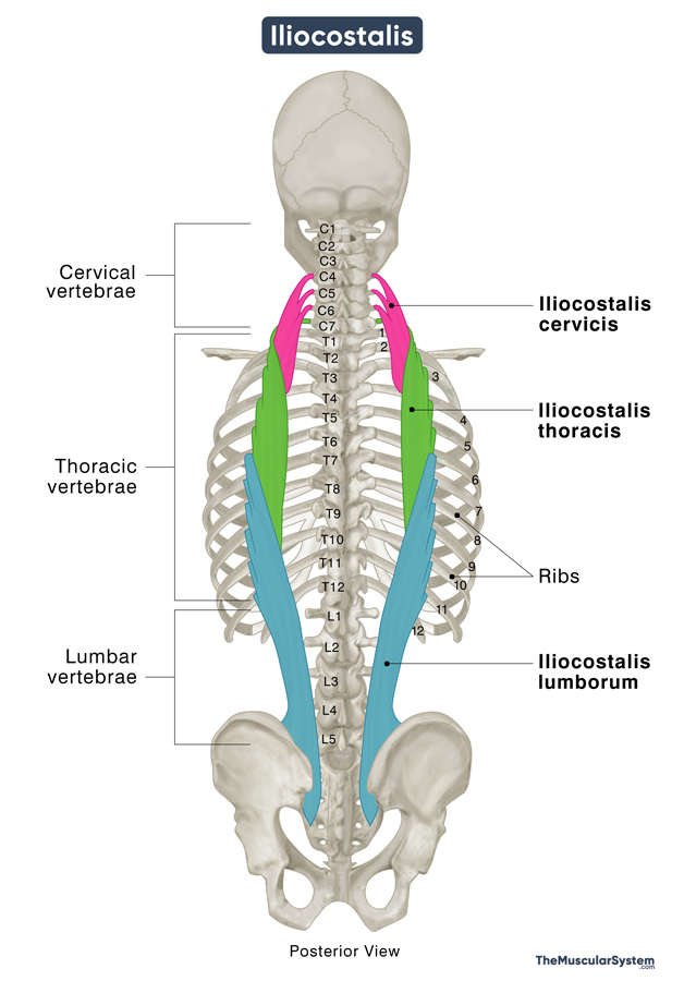
1. Iliocostalis Cervicis
The iliocostalis cervicis (also known as iliocostalis colli) is the uppermost part of the iliocostalis muscle, located at the back of the neck, with attachments to the cervical vertebrae. It originates from the superior borders of the angles of the 3rd to 6th ribs and inserts into the posterior tubercles of the transverse processes of the 4th to 6th cervical vertebrae (C4-C6).
2. Iliocostalis Thoracis
The middle portion of the muscle in the upper to middle back, the iliocostalis thoracis, originates as flat tendons from the upper borders of the angles of the 7th to 12th ribs. These tendons course upward, gradually becoming thicker and more muscular, before attaching to the upper edges of the 1st to 6th ribs and the transverse processes of the C7 vertebra (located at the base of the neck).
The origins of both the cervicis and thoracis portions are located medial to the insertion of the lumborum portion of the iliocostalis.
3. Iliocostalis Lumborum
It is the lowermost and biggest part of the iliocostalis muscle, located at the lower back region. It is further divided into the lumbar and thoracic (in the middle back) components.
The lumbar part (located in the lower back) starts at the lateral sacral crest, the inside edge of the hip bone (iliac crest), and the thoracolumbar fascia—a connective tissue in the lower back. From there, it extends upward and attaches to the transverse processes of the 1st through 4th lumbar vertebrae (L1-L4). Some part of it also inserts into the adjacent thoracolumbar fascia. The muscle fibers are arranged in layers, with the ones connected to L1 usually sitting closest to the surface and nearer the spine, while the fibers attached to L4 usually lie deeper and further out to the side.
The thoracic part (located in the mid-back region) originates from the same region as the lumbar part via flat ribbon-like tendinous sheaths that cover the lumbar part. From here, the muscle fibers course upwards and attach to the lower edges of the 5th to 12th ribs. The most lateral fibers attach to the highest rib (5th), while the most medial fibers attach to the lowest rib (12th).
Relations With Surrounding Muscles and Structures
The most lateral of the three erector spinae muscles, the iliocostalis, has the longissimus muscle lying medial to it. On the lateral side, it has the medial border of the scapula.
The cervicis part lies deep to the rhomboid and splenius capitis muscles. The upper part of the iliocostalis thoracis lies deep to the serratus posterior superior, while its lower part lies deep to the serratus posterior inferior.
Function
| Action | Unilateral: Bending the body trunk and neck sideways Bilateral: Extending or straightening the back and neck |
The iliocostalis works together with the other muscles of the erector spinae group.
Unilateral Contraction
When the muscle contracts unilaterally, it flexes the vertebral column on one side, causing the body to bend sideways. The iliocostalis lumborum and thoracis work on the lumbar and thoracic vertebrae to help bend the trunk of the body. Similarly, the cervicis part bends the neck and head by flexing the cervical vertebral joints.
Bilateral Contraction
When the muscle contracts bilaterally or on both sides, it extends the spinal column, straightening the trunk or bending it backward. Like during unilateral contraction, the thoracis and lumborum parts handle the thoracic and lumbar vertebrae, while the cervicis part works on the cervical vertebral joints to help extend the head and neck.
The muscle also plays an important role in maintaining good posture and keeping the spinal column stable even when it is not actively involved in its movements.
Innervation
| Nerve | Dorsal rami of spinal nerves |
The iliocostalis muscles are innervated by the lateral branches of the dorsal rami of spinal nerves, corresponding to the levels where the muscles are located along the spine.
- The cervicis part receives innervation from the lower cervical and upper thoracic nerves.
- The thoracis part, from the thoracic nerves.
- The lumborum part, from lower thoracic and lumbar nerves.
Blood Supply
The muscle has segmented blood supply with different arteries supplying to the 3 parts:
| Artery | Cervicis: Deep cervical, occipital, and vertebral arteries Thoracis: Dorsal or posterior branches of the posterior intercostal and subcostal arteries Lumborum: Lateral sacral arteries and the dorsal branches of the lumbar arteries |
References
- Iliocostalis: TeachMeAnatomy.info
- Iliocostalis Muscle: Kenhub.com
- Iliocostalis Colli Muscle: Elsevier.com
- Iliocostalis Thoracis Muscle: GetBodySmart.com
- Iliocostalis Thoracis Muscle: Elsevier.com
- Iliocostalis Lumborum Muscle: Elsevier.com
- Iliocostalis-Lumborum: HealthLine.com
Della Barnes, an MS Anatomy graduate, blends medical research with accessible writing, simplifying complex anatomy for a better understanding and appreciation of human anatomy.
- Latest Posts by Della Barnes, MS Anatomy
-
Tensor Tympani
- -
Stapedius
- -
Auricularis Posterior
- All Posts


