

Rectus Abdominis
By
Della Barnes, an MS Anatomy graduate, blends medical research with accessible writing, simplifying complex anatomy for a better understanding and appreciation of human anatomy.
Last updated:
24/05/2025Della Barnes, MS Anatomy
UX/UI Designer at - AdobeDella Barnes, an MS Anatomy graduate, blends medical research with accessible writing, simplifying complex anatomy for a better understanding and appreciation of human anatomy.
What is the Rectus Abdominis
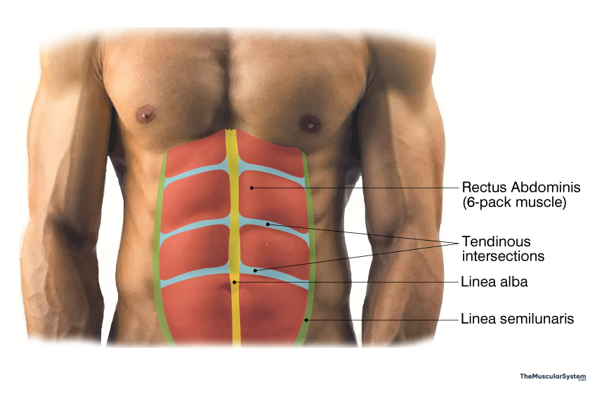
Rectus abdominis, referred to simply as the abs, is a long, paired, vertically oriented muscle located at the front of the abdomen, extending from the pelvic region to the ribs. It consists of two parallel muscle strips, separated by the linea alba, a fibrous band of connective tissue running down the midline of the abdomen. The muscle is segmented by tendinous intersections, which can create the appearance of a “six-pack” when body fat is low.
As one of the largest and most important muscles of the abdomen region, the rectus abdominis plays a key role in flexing the spine, stabilizing the trunk, and maintaining posture.
Anatomy
Location and Attachments
| Origin | Pubic crest, tubercle, and symphysis |
| Insertion | Xiphoid process of the sternum and the 5th to 7th costal cartilages |
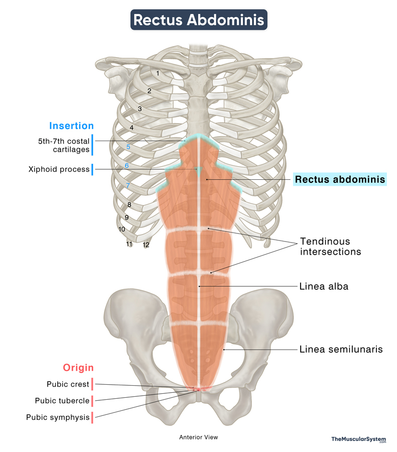
Origin
The rectus abdominis originates via two tendinous attachments. The larger lateral attachment arises from the pubic crest, pubic tubercle, and, occasionally, the pectineal line. The smaller medial attachment arises from the anterior surface of the pubic symphysis.
Insertion
The muscle fibers run vertically upward from the point of origin and insert via multiple muscular slips into the xiphoid process of the sternum and the costal cartilages of the 5th to 7th ribs. In some anatomical variations, the most laterally located fibers of the muscle may even extend as high as the 4th or even the 3rd rib. The medial slips primarily insert into the 6th and 7th costal cartilages and the lateral edges of the xiphoid process.
Structure
The rectus abdominis is enclosed by fibrous connective tissues that help define its borders and divisions. The linea alba, a thick aponeurotic band, runs along the midline of the abdomen, dividing the muscle into two parallel strips.
Additionally, three horizontal tendinous intersections segment the rectus abdominis at different levels:
- The lowest intersection is at the level of the umbilicus (belly button).
- The highest intersection is near the xiphoid process.
- The middle intersection lies between these two, approximately halfway along the muscle.
These intersections divide the muscle belly on each side into four distinct segments (eight in total), which can be visible, especially in individuals with a more athletic build, as the ‘six-pack.’
Relations With Surrounding Muscles and Structures
The rectus abdominis is the primary muscle forming the anterior abdominal wall, with a minor contribution from the pyramidalis muscle, which lies in front of its lower portion.
Laterally, the external oblique, internal oblique and transversus abdominis muscles flank the rectus abdominis. Their aponeuroses form the rectus sheath, a tough, multilayered fibrous compartment that encloses and supports the rectus abdominis. However, in the lower one-fourth of the muscle (below the arcuate line), the posterior wall of the sheath is absent, leaving the rectus abdominis to rest directly on the transversalis fascia.
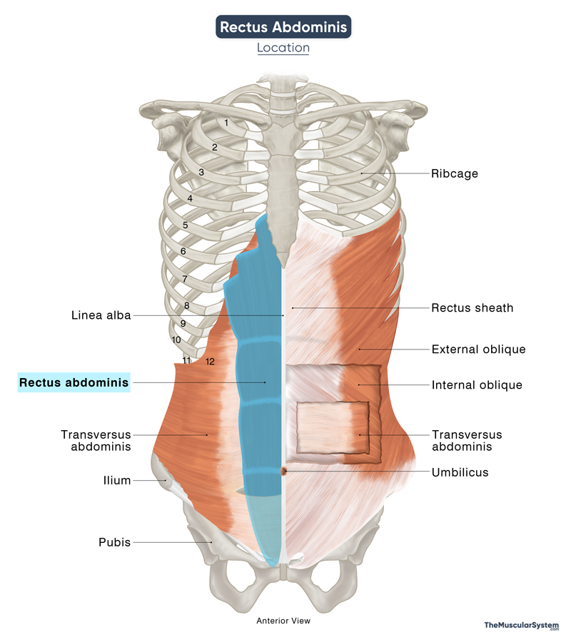
The linea semilunaris, a fibrous aponeurotic structure, marks the lateral borders of the rectus abdominis, separating it from the oblique muscles.
Function
| Action | Flexing the trunk at the lumbar spinal level Compressing the abdomen to help with bodily functions |
As a key core muscle, the rectus abdominis is involved in several important functions:
- Flexion of the Trunk: The muscle contracts to flex the lumbar spine and help bend the body forward, bringing the ribcage closer to the pelvis. The muscle is active during movements like sit-ups or when bending forward.
- Stabilization of the pelvis: By pulling the pubic bone upward, the rectus abdominis stabilizes the pelvis and prevents excessive arching of the lower back, reducing strain on the lumbar spine. This way, the muscle helps prevent the lower back from overextending during activities like squatting, running, or lifting weights.
- Intra-abdominal pressure regulation: The rectus abdominis works together with other abdominal muscles to compress the abdominal viscera (organs within the abdominal cavity), thereby increasing intra-abdominal pressure. It plays a role in forced expiration (pushing the diaphragm upward) and assists in urination, defecation, and labor.
- Core stability: The muscle supports the core by maintaining postural stability and reinforcing the abdominal wall to provide structural support to the abdominal contents and surrounding structures.
Antagonists
The spinal erectors or erector spinae group, consisting of the iliocostalis, longissimus, and spinalis, help straighten the back and bend it backward, making them antagonistic to the rectus abdominis.
Innervation
| Nerve | Anterior rami of T7 to T11 spinal nerves and subcostal nerve (T12) |
Both motor and sensory innervation come from the anterior or ventral rami of the lower thoracic spinal nerves — these include the T7 to T11 thoracoabdominal nerves and the T12 subcostal nerve.
These nerves travel between the internal oblique and transverse abdominis muscles before entering the rectus sheath to supply the muscle from its lateral aspect.
Blood Supply
| Artery | Inferior and superior epigastric arteries |
Primary blood supply comes from the Inferior and superior epigastric arteries, which also supply the umbilical and epigastric regions.
Additionally, the terminal branches of the T9 to T11 posterior intercostal arteries, the subcostal (T12) artery, and the deep circumflex iliac artery supply the muscle laterally and posteriorly.
References
- Rectus Abdominis Muscle: IMAIOS.com
- Rectus Abdominis: TeachMeAnatomy.info
- Rectus Abdominis Definition, Function & Location: Study.com
- Rectus abdominis muscle: Kenhub.com
- Arcuate Line: Osmosis.org
- Abdominal Muscles: ClevelandClinic.org
- Rectus Abdominis Muscle: GetBodySmart.com
- Rectus Abdominis Muscle: Elsevier.com
Della Barnes, an MS Anatomy graduate, blends medical research with accessible writing, simplifying complex anatomy for a better understanding and appreciation of human anatomy.
- Latest Posts by Della Barnes, MS Anatomy
-
Tensor Tympani
- -
Stapedius
- -
Auricularis Posterior
- All Posts


