Transversus Abdominis
Last updated:
26/05/2025Della Barnes, an MS Anatomy graduate, blends medical research with accessible writing, simplifying complex anatomy for a better understanding and appreciation of human anatomy.
What is the Transversus Abdominis
The transversus abdominis, also known as the transverse abdominal muscle, is a paired, flat, sheet-like muscle that forms the deepest layer of the lateral abdominal wall. It spans from the lower ribcage to the pelvis and wraps around the abdomen like a corset. It is the only abdominal muscle whose fibers run horizontally (transversely).
Together with other abdominal muscles, the transversus abdominis plays a crucial role in stabilizing the core, supporting abdominal organs, and maintaining intra-abdominal pressure, thereby contributing to overall trunk stability.
Anatomy
Location and Attachments
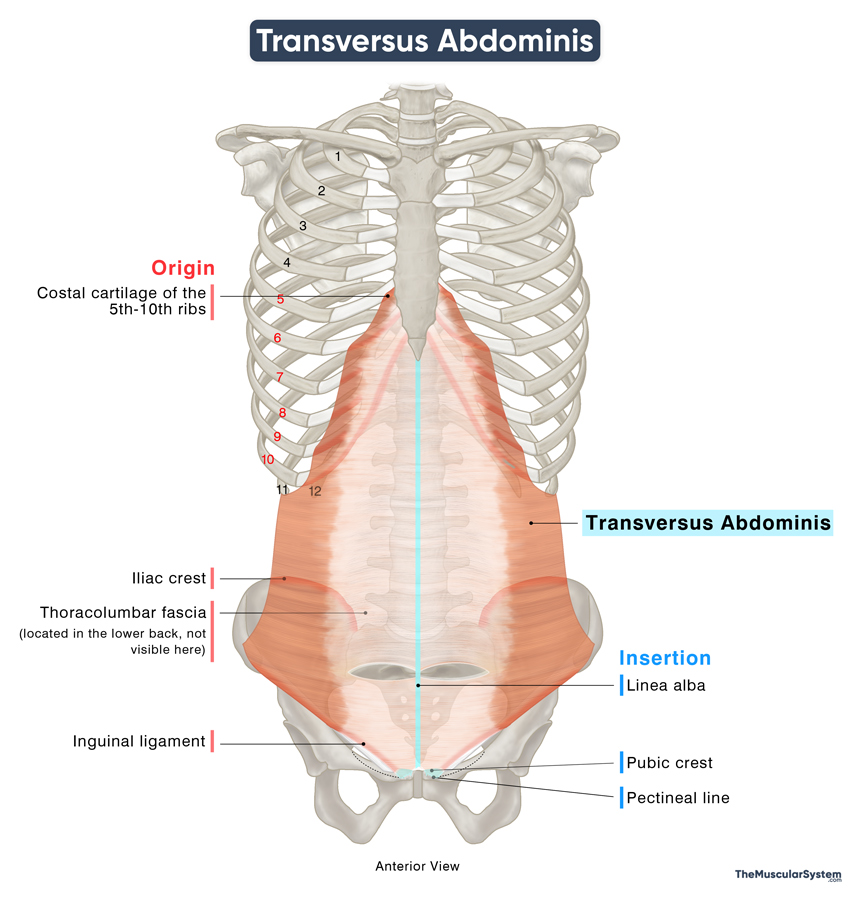
| Origin | Inguinal ligament, iliac crest, thoracolumbar fascia, and the costal cartilage of the 5th-10th ribs |
| Insertion | Linea alba, pubic crest, and the pectineal line of the pubis (via the conjoint tendon) |
Origin
The broad, flat muscle has several points of origin. It arises as multiple muscular slips from:
- The inner surfaces of the lower six costal cartilage, that is, from the 5th to 10th costal cartilage (the lowest two ribs do not have attached costal cartilage)
- The thoracolumbar fascia in the back
- The superior surface of the lateral one-third of the inguinal ligament
- The inner side of the lateral two-thirds of the iliac crest
Insertion
As indicated by its name, the muscle’s fibers run transversely in a medial and anterior direction. Like the two oblique muscles, its fibers turn aponeurotic, or flat and tendon-like, as they approach their insertion site.
The portion originating from the inguinal ligament runs horizontally and downward, forming the roof of the inguinal canal. Then, it merges with the aponeurotic fibers of the internal oblique muscle, forming the conjoint tendon. This tendon then attaches to the pubic crest and pectineal line (pecten pubis).
The remaining fibers from the superior part of the muscle travel anteriorly toward the rectus abdominis, contributing to the rectus sheath that covers it. Ultimately, the aponeurosis of the transversus abdominis inserts into the linea alba, the fibrous band extending along the midline of the abdomen.
Relations With Surrounding Muscles and Structures
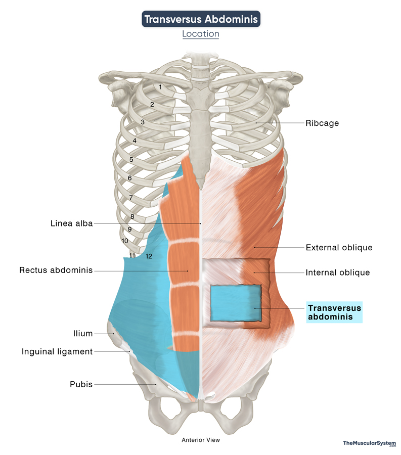
The transversus abdominis is the deepest of the anterolateral abdominal muscles. Positioned on the lateral side of the abdomen, it lies beneath both the internal and external oblique muscles. Medially, it is bordered by the rectus abdominis muscle.
Relation With the Rectus Sheath
This deep muscle plays an important role in forming the rectus sheath. In the upper abdomen, the aponeurosis (flat tendon) of the transversus abdominis passes behind the rectus abdominis, contributing to the posterior wall of the rectus sheath. The internal oblique is the only other muscle that assists in forming this portion of the sheath.
Approximately 2.5 cm below the umbilicus, a change occurs in the sheath’s structure. Here, the aponeuroses of the transversus abdominis, internal oblique, and external oblique all travel anterior to the rectus abdominis. This convergence creates the lower quarter of the anterior wall of the rectus sheath.
The point where the posterior wall ends is marked by a distinct anatomical landmark called the arcuate line. Below this line, the rectus abdominis rests directly on the transversalis fascia, as the posterior wall of the rectus sheath is absent.
Relation With the Transversalis Fascia
The transversalis fascia lies just below the lower free edge of the transversus abdominis muscle. This thin layer of connective tissue connects with nearby structures, such as the internal, external, and inguinal ligaments.
About 1 to 1.5 cm above the inguinal ligament, the transversalis fascia forms a small, round opening known as the deep inguinal ring. This ring is found roughly halfway between the anterior superior iliac spine (ASIS) and the pubic tubercle and serves as the entrance to the inguinal canal.
Sometimes, a slender band of muscle fibers from the transversus abdominis extends near the inner edge of the deep inguinal ring. This is called the interfoveolar ligament, which blends with the transversalis fascia, possibly helping reinforce this area.
Function
| Action | Compresses the abdomen and provides core stability |
Maintaining the structure of the abdominal wall
The transversus abdominis plays a crucial role in maintaining the structure of the abdominal wall. Working alongside the obliques and rectus abdominis, it helps preserve the shape and integrity of the torso. This deep muscle wraps around the sides of the body from front to back, forming a natural corset that supports and protects the abdominal organs, keeping them securely in place. When weakened, the transversus abdominis may contribute to an increased risk of abdominal hernias due to reduced internal support.
Role in intra-abdominal compression
In addition to structural support, the transversus abdominis is key in generating intra-abdominal compression. Along with other muscles in the lateral and anterior abdominal region, it helps compress the abdominal viscera—the internal organs within the abdominal cavity. This compression is essential for various bodily functions, including forced exhalation, defecation, and labor during childbirth.
Innervation
| Nerve | Thoracoabdominal nerves (T7-T11), subcostal nerve (T12), ilioinguinal nerve (L1), and iliohypogastric nerve (L1) |
The transversus abdominis gets its nerve supply mainly from the thoracoabdominal nerves, which branch out from the anterior rami of the 7th to 11th thoracic spinal nerves (T7-T11). Another key contributor is the subcostal nerve, coming from the 12th thoracic nerve (T12).
Furthermore, two nerves branching from the first lumbar spinal nerve (L1) — the iliohypogastric and ilioinguinal nerves — also innervate this muscle, particularly in the lower abdominal region.
Blood Supply
| Artery | Lower posterior intercostal arteries Subcostal artery Superficial circumflex iliac artery Deep circumflex iliac artery Superior epigastric artery Inferior epigastric artery |
As a flat abdominal muscle, the transversus abdominis receives its blood supply primarily from the lower posterior intercostal arteries (T9–T11) and the subcostal artery (T12).
The inferior portion of the muscle is supplied by the superficial and deep circumflex iliac arteries, as well as the superior and inferior epigastric arteries.
Additional vascular contributions arise from the lateral branches of the posterior lumbar arteries (L1–L4), which originate from the abdominal aorta.
References
- Anatomy, Abdomen and Pelvis: Abdominal Wall: NCBI.NLM.NIH.gov
- Transversus Abdominis Muscle: Elsevier.com
- Transversus Abdominis Muscle: Radiopaedia.org
- Transversus Abdominus Muscle | Function, Origin & Insertion: Study.com
- Transversus Abdominus Muscle: Kenhub.com
- Transversus Abdominus Muscle: IMAIOS.com
Della Barnes, an MS Anatomy graduate, blends medical research with accessible writing, simplifying complex anatomy for a better understanding and appreciation of human anatomy.
- Latest Posts by Della Barnes, MS Anatomy
-
Tensor Tympani
- -
Stapedius
- -
Auricular Muscles
- All Posts


