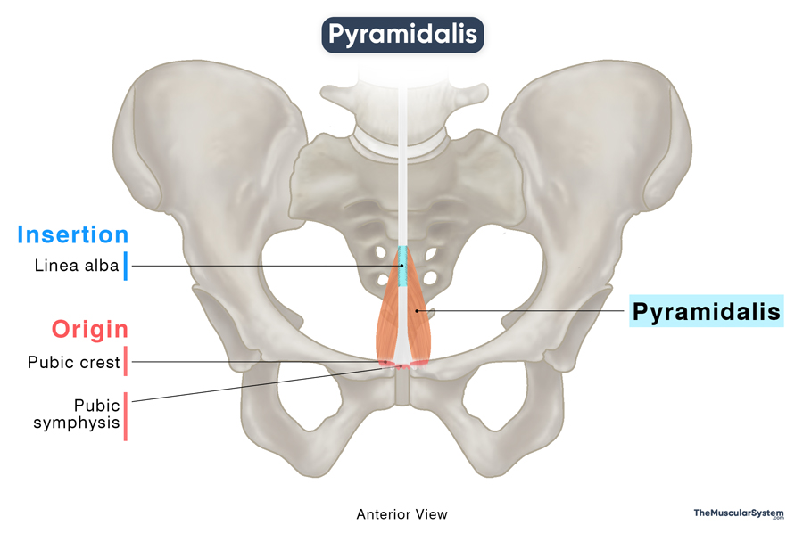
Pyramidalis
Last updated:
23/05/2025Della Barnes, an MS Anatomy graduate, blends medical research with accessible writing, simplifying complex anatomy for a better understanding and appreciation of human anatomy.
What is the Pyramidalis
The pyramidalis is a pair of small, triangular muscles in the anterior abdominal wall, specifically in the lower abdomen, in front of the inferior portion of the rectus abdominis (the “six-pack” muscle). It is the fifth and smallest muscle of the anterolateral abdominal wall, with the others being the external and internal obliques, transversus abdominis, and rectus abdominis.
The Pyramidalis muscle is present in only about 83% to 90% of people, regardless of gender. It may be present unilaterally or only on one side rather than as a pair, in rarer cases. It is usually considered to be a vestigial muscle and does not have any unique functions. When present, it helps with the actions of the other abdominal muscles.
Anatomy
Location and Attachments
| Origin | Pubic crest and pubic symphysis |
| Insertion | Linea alba |
Pyramidalis
Origin
The pyramidalis muscle arises from two points on the anterior part of the pubic bone. One portion originates from the pubic crest via tendinous fibers, while the other part arises from the anterior surface of the pubic symphysis through a fibrous attachment.
Insertion
The muscle fibers ascend medially from their origin and gradually taper into a small, triangular belly, giving them their characteristic triangular shape. The muscle inserts into the linea alba, the fibrous band running down the midline of the anterior abdominal wall. The insertion point is located approximately halfway between the pubic symphysis and the umbilicus.
Relations With Surrounding Muscles and Structures
Positioned, as earlier described, in front of the lower end of the rectus abdominis, the pyramidalis lies within the rectus sheath — a fibrous layered structure formed by the aponeurosis of the external and internal oblique and transversus abdominis. It is the most superficial muscle inside the sheath, located directly beneath the linea alba, a fibrous band running along the midline of the anterior abdominal wall.
Function
| Action | Tenses or tightens the linea alba |
When present, the pyramidalis muscle contracts to exert a slight pull on the linea alba, helping to tense and stabilize it. This action makes a minor contribution to the integrity and tautness of the lower abdominal wall. However, its role is considered functionally insignificant, as the primary abdominal muscles, such as the rectus abdominis and obliques, perform these functions effectively even in its absence.
Innervation
| Nerve | Subcostal nerve (T12) |
The muscle is innervated by the subcostal nerve, the anterior ramus of the twelfth thoracic spinal nerve (T12), which also innervates the adjacent rectus abdominis muscle.
Blood Supply
| Artery | Inferior epigastric artery |
The inferior epigastric artery gives off branches that supply the pyramidalis muscle with blood.
References
- Pyramidalis Muscle: Radiopaedia.org
- Pyramidalis Muscle: IMAIOS.com
- Pyramidalis Muscle: Kenhub.com
- Pyramidalis: TeachMeAnatomy.info
- Pyramidalis Muscle: Elsevier.com
- Pyramidalis Muscle (anatomy): GPnotebook.com
Della Barnes, an MS Anatomy graduate, blends medical research with accessible writing, simplifying complex anatomy for a better understanding and appreciation of human anatomy.
- Latest Posts by Della Barnes, MS Anatomy
-
Tensor Tympani
- -
Stapedius
- -
Auricularis Posterior
- All Posts


