Serratus Anterior
Last updated:
14/06/2024Della Barnes, an MS Anatomy graduate, blends medical research with accessible writing, simplifying complex anatomy for a better understanding and appreciation of human anatomy.
What is the Serratus Anterior
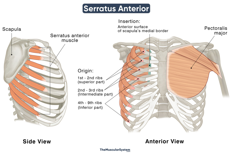
The serratus anterior is a large, thin fan-shaped sheet of muscle located at the sides and front of the thoracic wall. The muscle is easily visible, especially in people with an athletic build, along the sides of the ribs, beneath the axilla.
The large muscle is divided into three sections based on their insertion: the superior, intermediate, and inferior.
Anatomy
Location and Attachments
| Origin | Superior part: 1st and 2nd ribs Intermediate part: 2nd and 3rd ribs Inferior part: 4th to 8th ribs |
| Insertion | Superior part: Anterior surface of scapula’s superior angle Intermediate part: Anterior surface of scapula’s medial border Inferior part: Anterior surface of scapula’s inferior angle |
The muscle originates in finger-like fleshy strips (digitation) from the lateral and superior side of the upper 8 (sometimes 9) ribs. It gives the muscle a serrated or toothlike appearance, leading to the name ‘serratus’ anterior.
The points of origin spread towards the aponeuroses (connective tissue) surrounding the intercostals that run along the respective ribs. From the superior to inferior side, each of the 8-9 strips of tendon originate from their corresponding rib. The first is the only exception, as it arises from both the 1st and 2nd ribs.
After their origination, the muscle fibers run towards the posterior side of the body, along the side of the thoracic wall, to insert to the front of the scapula.
Relations With Surrounding Muscles and Structures
The serratus anterior lies deep to the pectoralis major and minor muscles, below the scapula. It can be palpated between the latissimus dorsi and pectoralis major.
The muscle is separated from the subscapularis by the supraserratus bursa, while the infraserratus bursa separates it from the ribs.
Functions
| Action | Abduction and upward rotation of the scapula when lifting the arm; stabilizing the scapula |
The muscle’s primary role is to control the movement of the scapula:
- Abducting the Scapula and Arm: Contraction of all three muscle parts makes the scapula move laterally and outward along the ribs. The resulting pull at the scapula’s inferior angle causes the shoulder joint’s upper part to shift, causing the arm to elevate. So, the muscle helps with lifting the arm overhead, above 90°.
- Rotating the scapula: Since the upper and lower parts of the muscle can contract separately, they help with the upward rotation of the scapula. When the upper serratus anterior contracts, it causes the scapula to rotate forward. In contrast, contraction of the lower part rotates the scapula back in place.
- Stabilizing the Scapula: During the above movement, the muscle’s superior part acts antagonistically by depressing the scapula. It keeps the scapula stable in its place along the chest wall.
- Lifting the Chest Wall: When the scapula stays fixed, the entire muscle works to elevate the ribs. All three heads work together in this, and since they are attached to the upper 8-9 ribs, their action helps lift the entire rib cage. This way, the serratus anterior helps with respiration and is considered an accessory inspiratory muscle.
The primary antagonists of the serratus anterior are the rhomboids major and minor and the trapezius muscles.
Innervation
| Nerve | Long thoracic nerve (C5, C6, and C7) |
The long thoracic nerve (C5-C7) is the primary and common source of innervation for the entire muscle. The superior and inferior parts have an additional innervation as well. An independent nerve branch with its roots in the C4, C5, and C6 innervates the superior part. Similarly, the inferior part receives nerve supply from an intercostal nerve branch.
Blood Supply
| Artery | Lateral and superior thoracic arteries, thoracodorsal artery |
The lateral and superior thoracic arteries branch from the axillary artery. The third source of blood supply, the thoracodorsal artery, is a branch of the subscapular artery.
References
- Anatomy, Thorax, Serratus Anterior Muscles: NCBI.NLM.NIH.gov
- What is the Serratus Anterior? – Definition & Function: Study.com
- Serratus Anterior Muscle: KenHub.com
- Serratus Anterior Muscle: IMAIOS.com
- Serratus Anterior: HealthLine.com
- Serratus Anterior Muscle: GetBodySmart.com
Della Barnes, an MS Anatomy graduate, blends medical research with accessible writing, simplifying complex anatomy for a better understanding and appreciation of human anatomy.
- Latest Posts by Della Barnes, MS Anatomy
-
Tensor Tympani
- -
Stapedius
- -
Auricularis Posterior
- All Posts


