Pectoralis Major
Last updated:
05/05/2023Della Barnes, an MS Anatomy graduate, blends medical research with accessible writing, simplifying complex anatomy for a better understanding and appreciation of human anatomy.
What is the Pectoralis Major
The pectoralis major is a large, triangular, or fan-shaped superficial muscle lying at the anteriormost position in the chest cavity. It forms the front wall of the axilla, making up most of the chest and breast. The paired muscle is often called the ‘pecs’ in combination with the smaller muscle pectoralis minor.
Anatomy
Location and Attachments
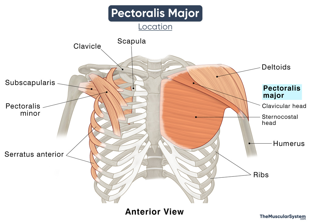
Since it has a broad origin, the muscle is divided into 2 heads, the clavicular and the sternocostal heads, named after their points of origin.
| Origin | Clavicular head: Medial half of the clavicle, on the anterior side Sternocostal head: Anterior side of the clavicle, costal cartilages 1-6, and the external oblique aponeurosis |
| Insertion | Lateral lip of the bicipital (intertubercular) groove of the humerus and the greater tubercle’s crest |
The muscle may also have a smaller third head, the abdominal head. It arises from the tendon sheath of the external oblique muscle, having the same insertion point as the other two heads. This head often varies from large to small or even completely absent.
After originating from the anterior side of the clavicle, all the heads run laterally, coming together to form a large tendon which then inserts into the greater tubercle crest.
Relations to Other Muscular Structures
As mentioned above, the pectoralis major is the most superficial muscle in the chest. These two muscles are much more visible in males than females because they are covered with breast tissues. In males, a deep layer of thoracic fascia, subcutaneous tissues, and skin cover them.
The muscle lies on top of the muscles serratus anterior and pectoralis minor, covering the ribs 1-6 at the front. The pectoralis major, along with the deltoids and the clavicle, forms a triangular depression referred to as the infra-clavicular fossa. This fossa is an important landmark in surgeries involving the subclavian artery.
One unique thing about the pectoralis major is that the length of its muscle fibers may vary between individuals. In most muscles in the human body, the muscle fibers maintain uniform length in everyone. The differing length of the pectoralis major fibers allow for variation in the power production due to differences in the degree of contraction (muscle shortening velocities).
Functions
| Action | Forward flexion, adduction of the arm over and across the chest, and rotating the arm at the shoulder; abduction and depression of the scapula (secondary action). |
- It is one of the primary muscles for the adduction of the arm, especially when working together with the latissimus dorsi. This action is used most when doing pull-ups or rock climbing.
- The pectoralis major also helps with the medial or internal rotation of the arm at the shoulder joint.
- The clavicular head helps flex the arm at the shoulder joint from its resting position. As an antagonist to this movement, the sternocostal head helps extend the arm back to its resting position to the side of the body.
- With its fixed attachment at the greater tubercle of the humerus, it can help elevate the thorax during inspiration or inhalation. It is a minor function that may be particularly useful during forced breathing, as is some physical distress.
The primary antagonists of the pectoralis major in the neutral position are the deltoids (lateral and posterior parts).
Its agonists are the teres minor, subscapularis, and the latissimus dorsi (partially).
Innervation
| Nerve | Clavicular head: Lateral pectoral nerve (C5, and C6) Sternocostal head: Medial pectoral nerve (C7, C8, and T1) |
Both the lateral and medial pectoral nerves branch out from the brachial plexus.
Blood Supply
| Artery | Pectoral branch of the thoracoacromial artery |
Apart from the thoracoacromial artery, the perforating branches of the internal thoracic artery also supply to the pectoralis major.
The pectoral vein drains the muscle, draining directly into the subclavian vein.
References
- Pectoralis Major Muscle – Attachment, Action & Innervation: GetBodySmart.com
- Pectoralis Major Muscle: RadioPaedia.org
- Pectoralis Major: Rad.Washington.edu
- Pectoralis Major Muscle: KenHub.com
- A Detailed Review on the Clinical Anatomy of the Pectoralis Major Muscle: Core.AC.UK
- Muscles of the Pectoral Region: TeachMeAnatomy.info
- Pectoralis Major (Anatomy): PrimaryCareNotebook.com
- Abdominal Head of Pectoralis Major Muscle: InnerBody.com
Della Barnes, an MS Anatomy graduate, blends medical research with accessible writing, simplifying complex anatomy for a better understanding and appreciation of human anatomy.
- Latest Posts by Della Barnes, MS Anatomy
-
Temporoparietalis
- -
Corrugator Supercilii
- -
Depressor Supercilii
- All Posts


