Teres Major
Last updated:
05/05/2023Della Barnes, an MS Anatomy graduate, blends medical research with accessible writing, simplifying complex anatomy for a better understanding and appreciation of human anatomy.
What is the Teres Major
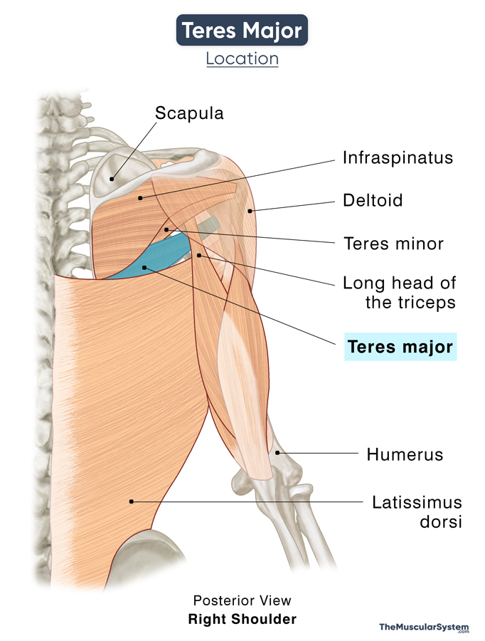
The teres major is a rectangular, thick, flat shoulder muscle extending from the lower scapular region below the armpit to the upper (proximal) part of the humerus’s shaft.
It is one of the 7 scapulohumeral muscles that attach the humerus to the scapula, connecting the arm to the shoulder. The muscle works with the back muscle latissimus dorsi to move and rotate the arm at the shoulder or glenohumeral joint.
Anatomy
Location and Attachments
It is located deep to or beneath the deltoids in the back of the shoulder. Teres major, along with the latissimus dorsi and subscapularis, form the back wall of the axilla (the region where the arm joins with the shoulder).
| Origin | The back surface of the scapula’s inferior angle |
| Insertion | The medial lip of the bicipital groove (intertubercular sulcus) on the anterior side of the humerus |
Relations to Other Muscular Structures
The teres major lies just below the teres minor, close enough that their bellies and tendons may somewhat blend. Due to their similar names and proximity, the teres major is often thought to be a rotator cuff muscle like the teres minor. However, unlike the latter, it does not attach to the glenohumeral joint capsule. It thus is not considered a rotator cuff muscle.
The back surface of the muscle at its origin at the inferior angle of the scapula remains covered under the infraspinatus and teres minor muscles. From its origin, it runs across the front of the long head of the triceps to insert into the humerus. This end lies close to the subscapularis muscle.

The lower margin of this muscle runs parallel to the upper margin of the latissimus dorsi, close enough that their adjacent muscle fibers may sometimes even fuse.
Functions
| Action | Adduction, extension, and medial rotation of the humerus at the shoulder joint. |
The teres major is primarily involved in the movement of the humerus, thus, the upper arm, at the shoulder joint. Its functions are:
- It assists in rotating the humerus towards the body, for example, when crossing your arms across the chest.
- The muscle adducts the arm or brings it back towards the body’s midline from an extended position.
- When its point of attachment to the humerus is fixed, the teres major assists the latissimus dorsi and pectoralis major to move the body closer to the arm superiorly. For example, doing pull-ups or climbing rocks; this is why the muscle is sometimes referred to as a ‘climbing muscle.’
- It also helps extend the arm and raise it towards the back, like when you are pitching a baseball or bringing the arms downward in the back during arm circling.
- Being one of the scapulohumeral muscles, it helps keep the humerus head in place in the glenohumeral joint, stabilizing it as well.
Innervation
| Nerve | Lower subscapular nerve (nerve roots C5, C6, C7) |
The lower subscapular nerve that supplies the teres major is a branch of the posterior cord of the brachial plexus.
Blood Supply
| Artery | The subscapular artery (thoracodorsal branch) and the posterior circumflex humeral artery |
References
- Teres Major: Origin, Action & Insertion: Study.com
- Teres Major Muscle: KenHub.com
- Teres Major Muscle – Attachments, Action & Innervation: GetBodySmart.com
- Teres Major: HealthLine.com
- Teres Major Muscle: RadioPaedia.org
- Anatomy, Shoulder and Upper Limb, Teres Major Muscle: NCBI.NLM.NIH.gov
- Teres Major: Rad.Washington.edu
Della Barnes, an MS Anatomy graduate, blends medical research with accessible writing, simplifying complex anatomy for a better understanding and appreciation of human anatomy.
- Latest Posts by Della Barnes, MS Anatomy
-
Tensor Tympani
- -
Stapedius
- -
Auricular Muscles
- All Posts


