

Quadratus Plantae
By
Della Barnes, an MS Anatomy graduate, blends medical research with accessible writing, simplifying complex anatomy for a better understanding and appreciation of human anatomy.
Last updated:
29/10/2025Della Barnes, MS Anatomy
UX/UI Designer at - AdobeDella Barnes, an MS Anatomy graduate, blends medical research with accessible writing, simplifying complex anatomy for a better understanding and appreciation of human anatomy.
What is the Quadratus Plantae
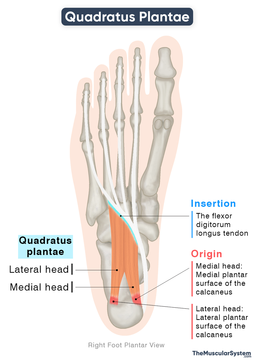
The quadratus plantae, also known as the flexor accessorius, is a short, two-headed, intrinsic muscle located along the center of the plantar surface of the foot. It forms part of the second layer of plantar muscles, along with the lumbricals.
The quadratus plantae assists in flexing the lateral toes, helping to stabilize the foot during walking and other movements.
Anatomy
The two heads of the muscle arise from different parts of the calcaneus or heel bone, and are separated by the long plantar ligament.
Location and Attachments
| Origin | — Medial head: Medial side of the plantar surface of the calcaneus bone — Lateral head: Lateral side of the plantar surface of the calcaneus bone |
| Insertion | The flexor digitorum longus tendon |
Origin
The medial head is larger and more muscular. It originates from the concave medial portion of the calcaneus on its plantar (inferior) surface, just below the groove that holds the tendon of the flexor hallucis longus as it passes toward the sole.
The lateral head is smaller and more tendinous. It arises from the lateral aspect of the plantar surface of the calcaneus, anterior to the lateral tubercle of the calcaneal tuberosity, with additional fibers often originating from the long plantar ligament.
Insertion
Both heads course downward and unite to form a thick tendinous band that inserts into the tendon of the flexor digitorum longus, near the point where it divides into four tendons for the lateral toes. Through this insertion, the quadratus plantae can send tendinous slips into each of these four divisions.
Variations
The muscle displays several variations where it only sends its slips to the 2nd to 3rd or 4th digits. Sometimes, the lateral head may be much smaller or missing, while in some cases, the muscle may be absent altogether.
Relations With Surrounding Muscles and Structures
The quadratus plantae lies in the second layer of the sole, deep to the flexor digitorum brevis and abductor digiti minimi form the first layer, and superficial to the adductor hallucis from the third layer.
It lies lateral to the abductor hallucis muscle and the tendon of the flexor hallucis longus.
The lateral plantar nerve and vessels pass along its superficial surface, separating it from the first layer, while the medial plantar nerve and vessels lie on its medial side.
Function
| Action | Assisting the flexor digitorum longus in flexing the 2nd to 5th toes |
The quadratus plantae works closely with the flexor digitorum longus (FDL). Its main role is to pull the FDL tendons toward the calcaneus, straightening their line of pull. This makes it easier for the FDL to bend the toes straight and have a better grip.
During walking, especially when the foot pushes off the ground, the FDL alone cannot fully flex the toes because it is already fully contracted in plantar flexion. The quadratus plantae assists by tightening the long flexor tendons even more, allowing the toes to bend and help propel the body forward.
By acting on the tendons of the FDL, the quadratus plantae aids in flexion at all the joints of the four smaller toes: the metatarsophalangeal, proximal interphalangeal, and distal interphalangeal joints.
It is one of the few muscles that are unique to the foot, with no equivalent in the hand.
Antagonists
It has no direct antagonists, but since it assists the flexor digitorum longus in flexing the lateral toes, the extensor digitorum longus and extensor digitorum brevis, which extend these toes, act as its functional antagonists.
Innervation
| Nerve | Lateral plantar nerve (S1-S3) |
One of the two terminal branches of the tibial nerve, the lateral plantar nerve, innervates this muscle. It carries fibers from the first to third sacral nerve roots (S1-S3).
Blood Supply
| Artery | Medial and lateral plantar arteries |
Blood supply to this muscle comes from the medial and lateral plantar arteries, both branches of the posterior tibial artery. The deep plantar arterial arch, a branch of the lateral plantar artery, also supplies the muscle.
References
- Quadratus Plantae: TeachMeAnatomy.info
- Quadratus Plantae Muscle: Kenhub.com
- Quadratus Plantae Muscle: Elsevier.com
- Quadratus Plantae Muscle: IMAIOS.com
Della Barnes, an MS Anatomy graduate, blends medical research with accessible writing, simplifying complex anatomy for a better understanding and appreciation of human anatomy.
- Latest Posts by Della Barnes, MS Anatomy
-
Tensor Tympani
- -
Stapedius
- -
Auricularis Posterior
- All Posts


