

Extensor Digitorum Longus
By
Della Barnes, an MS Anatomy graduate, blends medical research with accessible writing, simplifying complex anatomy for a better understanding and appreciation of human anatomy.
Last updated:
04/09/2025Della Barnes, MS Anatomy
UX/UI Designer at - AdobeDella Barnes, an MS Anatomy graduate, blends medical research with accessible writing, simplifying complex anatomy for a better understanding and appreciation of human anatomy.
The extensor digitorum longus is a long, narrow, unipennate muscle located on the anterior side of the front of the lower leg, extending down to the toes. It is one of the four muscles in the anterior compartment of the lower leg, along with the tibialis anterior, extensor hallucis longus, and fibularis tertius muscles.
It contributes to foot and leg movements by extending the toes and assisting in dorsiflexion of the foot.
Anatomy
Location and Attachments
| Origin | Lateral condyle of the tibia, the upper three-fourths of the anterior side of the fibular body, and the adjacent interosseous membrane |
| Insertion | Middle and distal phalanges of the 2nd to 5th toes |
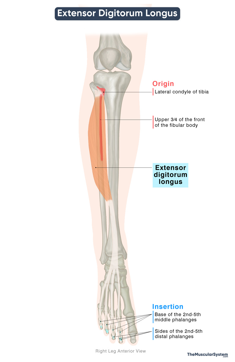
Origin
The muscle begins from several places in the upper part of the leg, just below the knee, including:
- The lower surface of the lateral side of the lateral condyle of the tibia.
- The upper three-fourths of the inner surface of the fibular body. It is the largest point of origin for its tendons.
- The front of the adjacent interosseous membrane between the tibia and fibula.
Extensor Digitorum Longus Insertion
From its origin, the muscle runs downward along the front of the leg, forming the fleshy muscle belly. Just above the ankle, it narrows into a tendon that passes under the superior and inferior extensor retinacula (fibrous bands that keep the tendons entering the foot in place).
Division into 4 tendons
As it comes out of the inferior extensor retinaculum, the single tendon divides into four smaller tendons, each going to one of the four lateral toes, the 2nd to 5th. These tendons share a synovial sheath that helps them glide smoothly over the dorsum or top of the foot.
Formation of the extensor hood
As the tendons cross over the proximal phalanges of their respective toes, each one flattens into a triangular sheet called an extensor hood. The medial part of each hood blends with fibers from the lumbrical and interossei muscles, while the lateral parts of toes 2-4 also receive fibers from the extensor digitorum brevis muscle.
Formation of the smaller inserting bands
With these contributions, each of the four tendons forms three fibrous bundles that insert into different points:
- The central bundle, or band, inserts into the base of the middle phalanx of the 2nd to 5th digits.
- The two lateral bands go further along the sides of each toe to insert at the base of the distal phalanges.
Relations With Surrounding Muscles and Structures
The extensor digitorum longus is the most lateral muscle in the extensor compartment of the leg. On its inner side lie the tibialis anterior and the extensor hallucis longus. Between it and the tibialis anterior pass the anterior tibial vessels and the deep fibular nerve.
At the level of the ankle, within the inferior extensor retinaculum, the tendon of extensor digitorum longus is positioned between the fibularis tertius tendon laterally and the extensor hallucis longus tendon medially.
Its tendons then approach the toes as they continue across the dorsum of the foot, where they lie superficial to the extensor digitorum brevis muscle.
Function
| Action | Extension of the 2nd to 5th digits at the metatarsophalangeal joints, and dorsiflexion of the foot |
Toe extension
The main function of the extensor digitorum longus is to extend the 2nd to 5th toes. Even though its tendons reach the middle and distal phalanges, the direction of the pull of this muscle is such that, by itself, it can only extend the toes at the metatarsophalangeal joints.
When it works together with the lumbricals, the extensor digitorum longus also helps extend the interphalangeal joints. In this way, both muscle groups act together to produce smooth extension of the entire length of the toes.
Dorsiflexion of the foot
It works in synergy with the other three muscles in the anterior compartment of the lower leg to dorsiflex the foot at the ankle joint. It means bringing the top of the foot toward the shin.
If the foot is planted on the ground, the muscle helps pull the leg and trunk forward over the foot, shifting the body’s weight from the heel toward the toes as happens in walking.
Both of these actions are vital for the gait cycle. Another minor function of the muscle is assisting with eversion of the foot.
Antagonists
The flexor digitorum longus, a muscle in the deep posterior compartment of the leg, and the flexor digitorum brevis, located in the foot, both flex the four lateral toes. Since they perform the opposite movement to the extensor digitorum longus, they act as its antagonists.
Innervation
| Nerve | Deep fibular nerve (L5, S1) |
The primary innervation of the muscle comes from the deep fibular nerve, arising from the fifth lumbar and first sacral nerve roots (L5-S1). It is a branch of the common fibular nerve, which is one of the two primary branches of the sciatic nerve.
Blood Supply
| Artery | Anterior tibial artery |
The Extensor Digitorum Longus muscle mainly receives its blood supply from the anterior tibial artery, a branch of the popliteal artery. The distal portion of the muscle receives additional vasculature from the perforating branches of the fibular artery.
References
- Extensor Digitorum Longus: Elsevier.com
- Extensor Digitorum Longus Muscle: Kenhub.com
- Extensor Digitorum Longus: TeachMeAnatomy.info
- Extensor Digitorum Longus Muscle: Radiopaedia.org
- Extensor Digitorum Longus Muscle | Anatomy, Action & Pain: Study.com
Della Barnes, an MS Anatomy graduate, blends medical research with accessible writing, simplifying complex anatomy for a better understanding and appreciation of human anatomy.
- Latest Posts by Della Barnes, MS Anatomy
-
Tensor Tympani
- -
Stapedius
- -
Auricularis Posterior
- All Posts


