

Anterior Compartment of the Leg
By
Della Barnes, an MS Anatomy graduate, blends medical research with accessible writing, simplifying complex anatomy for a better understanding and appreciation of human anatomy.
Last updated:
12/09/2025Della Barnes, MS Anatomy
UX/UI Designer at - AdobeDella Barnes, an MS Anatomy graduate, blends medical research with accessible writing, simplifying complex anatomy for a better understanding and appreciation of human anatomy.
The anterior compartment of the leg is one of the four fascial compartments of the lower limb. It occupies the front of the leg, extending from the knee to the ankle and continuing onto the dorsum of the foot. This compartment contains four muscles.
Functionally, the muscles of this compartment act mainly to dorsiflex the ankle, which means pulling the foot upward toward the shin. This movement lifts the foot during the swing phase of walking and keeps it steady as the heel makes contact with the ground.
The compartment is bounded on all sides by distinct structures:
- Anteriorly, by the deep fascia of the leg (crural fascia).
- Medially, by the lateral surface of the tibia.
- Laterally, by the anterior intermuscular septum, which separates it from the lateral compartment.
- Posteriorly, by the interosseous membrane between the tibia and fibula, which separates it from the deep posterior compartment.
Muscles in the Anterior Compartment and Their Anatomy
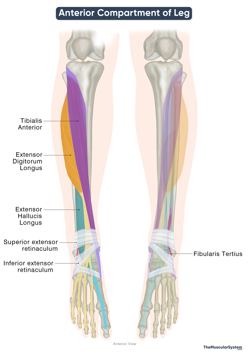
Here are the muscles in the anterior compartment, each with distinct attachments in the foot that allow them to perform specialized functions alongside their common role in dorsiflexion.
| Name | Origin | Insertion | Function |
|---|---|---|---|
| Tibialis Anterior | Lateral surface of upper tibia, adjacent interosseous membrane | Medial & inferior surfaces of medial cuneiform, base of 1st metatarsal | Dorsiflexion at the ankle, inversion of the foot |
| Extensor Hallucis Longus | Middle anterior fibula, adjacent interosseous membrane | Base of distal phalanx of hallux (big toe) | Extension of the great toe, dorsiflexion at the ankle |
| Extensor Digitorum Longus | Lateral condyle of tibia, upper ¾ of anterior fibula, adjacent interosseous membrane | Middle & distal phalanges of 2nd–5th toes | Extension of the 2nd–5th toes at the MTP joints, dorsiflexion at the ankle |
| Fibularis Tertius | Distal ⅓ of medial fibula, adjacent interosseous membrane | Dorsal base of 5th metatarsal | Dorsiflexion and eversion of the foot at the ankle |
All four tendons of the anterior compartment pass deep to the superior and inferior extensor retinacula, which hold them in place and prevent bowstringing during dorsiflexion.
Being in the same compartment, they share a common neurovascular supply:
Innervation: They receive motor supply from the deep fibular nerve, which carries fibers from the L4, L5, and S1 nerve roots. The deep fibular nerve arises from the common fibular nerve, itself a terminal branch of the sciatic nerve.
Blood supply: Vascularization is provided mainly by the anterior tibial artery, a continuation of the popliteal artery.
References
- Anterior Compartment of the Leg: Radiopaedia.org
- Anatomy, Bony Pelvis and Lower Limb: Leg Anterior Compartment: NCBI.NLM.NIH.gov
- Anterior Muscles of the Leg: Kenhub.com
- Muscles in the Anterior Compartment of the Leg: TeachMeAnatomy.info
Della Barnes, an MS Anatomy graduate, blends medical research with accessible writing, simplifying complex anatomy for a better understanding and appreciation of human anatomy.
- Latest Posts by Della Barnes, MS Anatomy
-
Tensor Tympani
- -
Stapedius
- -
Auricularis Posterior
- All Posts


