

Tibialis Anterior
By
Della Barnes, an MS Anatomy graduate, blends medical research with accessible writing, simplifying complex anatomy for a better understanding and appreciation of human anatomy.
Last updated:
08/10/2025Della Barnes, MS Anatomy
UX/UI Designer at - AdobeDella Barnes, an MS Anatomy graduate, blends medical research with accessible writing, simplifying complex anatomy for a better understanding and appreciation of human anatomy.
What is the Tibialis Anterior
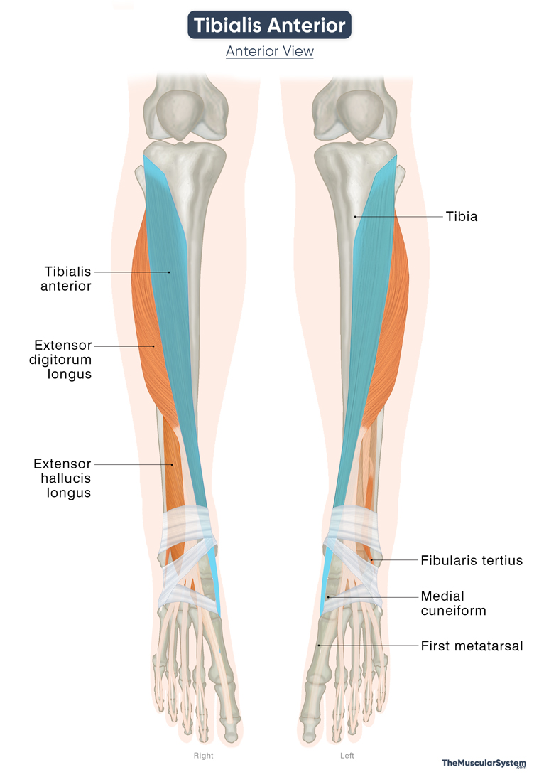
The tibialis anterior, also known as tibialis anticus, is one of the largest and strongest muscles of the front of the lower leg, lying beside the tibia or shinbone. It is a long, narrow, fusiform muscle that occupies the anterior compartment of the leg along with the extensor hallucis longus, extensor digitorum longus, and fibularis tertius.
It is one of the primary dorsiflexors of the human leg and plays a vital role in the gait cycle. The muscle also assists in inverting the foot and maintaining balance.
Anatomy
Location and Attachments
| Origin | Lateral surface of the upper part of the tibia and the adjacent interosseous membrane |
| Insertion | Medial and inferior surfaces of the medial cuneiform, and the base of the first metatarsal |
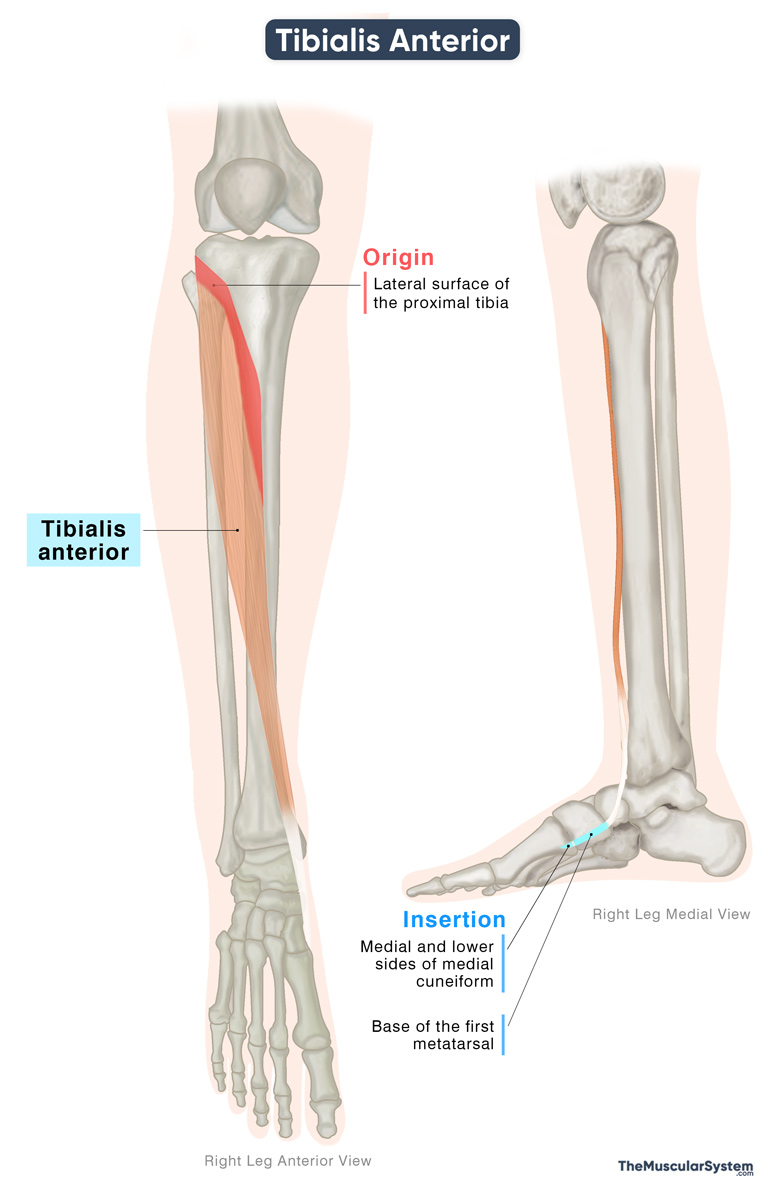
Origin
The muscle originates from several points on the proximal tibia and the surrounding fascial structures, including:
- The lateral condyle of the tibia.
- The lateral surface of the upper half or two-thirds of the tibia.
- The anterior side of the interosseous membrane that separates the tibia and fibula bones.
- The deep surface of the fascia cruris, also called the deep fascia of the leg.
- The anterior side of the intermuscular septum that separates this muscle from the extensor digitorum longus in the anterior compartment.
Insertion
After arising from its points of origin, the muscle fibers come together to form a fleshy belly that gradually tapers into a long tendon in the lower third of the tibia. This tendon passes down the leg, crosses the ankle joint, and runs along the dorsum or upper surface of the foot toward the midfoot region.
It finally inserts into the medial and bottom (plantar) surfaces of the medial cuneiform bone, with some fibers also attaching to the base of the first metatarsal bone.
Relations With Surrounding Muscles and Structures
Tibialis anterior is the most medial muscle in the anterior compartment of the leg, and its tendon remains the most medial as it passes across the ankle and into the foot.
The muscle lies just under the skin and fascia, resting directly on the tibia and the attached interosseous membrane. On its lateral side, it is bordered by the extensor digitorum longus and the extensor hallucis longus.
The fleshy belly of the muscle covers the anterior tibial artery and the deep fibular nerve, both major neurovascular structures that supply this compartment.
Tendon Pathway
On the distal side, the tibialis anterior tendon passes beneath the superior and inferior extensor retinacula at the front of the ankle. These fibrous bands hold the extensor tendons in place during ankle and foot movements. Unlike the other tendons here, the tibialis anterior tendon has its own synovial sheath. This sheath begins just above the superior extensor retinaculum and extends to the talonavicular joint, allowing the tendon to glide smoothly without friction.
Function
| Action | Dorsiflexion at the ankle joint and inversion of the foot |
Since the tibialis anterior tendon crosses the ankle, subtalar, and midtarsal joints, it acts on them all, helping with moving the foot in different directions.
Dorsiflexion
The tibialis anterior is the strongest muscle that performs dorsiflexion of the foot at the ankle joint. Dorsiflexion means pulling the foot upward toward the shin, and this movement is crucial for walking because it lifts the foot during the swing phase of gait, keeping it from dragging.
Inversion
The tibialis anterior also turns the sole of the foot inward, a movement known as inversion. This occurs because the muscle begins on the outer side of the tibia, but its tendon attaches on the inner side of the foot. So, when it contracts, it pulls up the bones of the inner arch, specifically the medial cuneiform, first metatarsal, navicular, and talus, into an inward, supinated position. It is similar to pulling on a rope that’s tied to the inner side of a shoe. It will change the position of the entire shoe.
Most of this inward turning happens at two joints just below the ankle: the subtalar joint (between the talus and calcaneus) and the midtarsal joint (between the talus and navicular).
Since the tibialis anterior is such a strong inverter, the muscles on the outer side of the leg often contract at the same time to pull the foot outward in a movement called eversion. This balances the movement, allowing dorsiflexion to happen without excessive inversion.
Stability
The muscle also supports the medial longitudinal arch of the foot, which is higher than the lateral arch. It is made up of the calcaneus, talus, navicular, the three cuneiforms, and the first three metatarsals. Keeping this arch lifted helps with balance, stability, and sharing body weight evenly when standing or walking.
Antagonists
As the main dorsiflexor of the ankle, its antagonists are the plantarflexor muscles of the posterior compartment of the leg, including the gastrocnemius, soleus, plantaris, and tibialis posterior.
In addition, because tibialis anterior also produces inversion of the foot, the evertor muscles of the lateral compartment, primarily the fibularis longus and fibularis brevis, are antagonistic to it.
Innervation
| Nerve | Deep fibular nerve (L4-L5) |
It is innervated by the deep fibular nerve, originating from the 4th and 5th lumbar nerve branches. It is a branch of the common fibular nerve, which itself branches from the sciatic nerve.
Blood Supply
| Artery | Anterior tibial artery |
The primary blood supply to the Tibialis Anterior muscle comes from the anterior tibial artery, which supplies the proximal part, or the muscle belly. It is one of the terminal branches of the popliteal artery.
The distal tendon part of the muscle receives blood supply specifically from the medial tarsal arteries, which branch off the dorsalis pedis artery, a continuation of the anterior tibial artery.
Additional vascular supply may come from branches of the posterior tibial artery.
References
- Tibialis Anterior Muscle: Elsevier.com
- Anatomy, Bony Pelvis and Lower Limb: Tibialis Anterior Muscles: NCBI.NLM.NIH.gov
- Tibialis Anterior: Rad.UW.edu
- Tibialis Anterior Muscle: Kenhub.com
- Tibialis Anterior Muscle – Attachments, Actions & Innervation: GetBodySmart.com
- Tibialis Anterior Muscle: Radiopaedia.org
- Tibialis Anterior: TeachMeAnatomy.info
Della Barnes, an MS Anatomy graduate, blends medical research with accessible writing, simplifying complex anatomy for a better understanding and appreciation of human anatomy.
- Latest Posts by Della Barnes, MS Anatomy
-
Tensor Tympani
- -
Stapedius
- -
Auricularis Posterior
- All Posts


