Fibularis Brevis
Last updated:
10/09/2025Della Barnes, an MS Anatomy graduate, blends medical research with accessible writing, simplifying complex anatomy for a better understanding and appreciation of human anatomy.
What is the Fibularis Brevis
The fibularis brevis, also known as the peroneus brevis, is a short, slender muscle situated on the lateral side of the lower leg, running from the lower portion of the fibula to the sole of the foot. It lies deep to the fibularis longus, and together they occupy the lateral compartment of the leg. It is also one of the three muscles in the fibularis group.
Its primary function is eversion of the foot, while it also assists with plantarflexion of the ankle. These actions are essential for maintaining balance and providing lateral stability during the gait cycle.
Anatomy
Location and Attachments
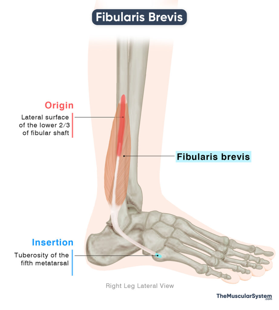
| Origin | Lateral surface of the lower two-thirds of the fibular shaft |
| Insertion | The tuberosity on the lateral side of the fifth metatarsal base |
Origin
The Fibularis Brevis muscle primarily arises from the distal side of the fibula, specifically from the lateral side of its body or shaft. Some part of the muscle also originates from the adjacent intermuscular septa that separate the anterior and posterior compartments of the lower leg.
Insertion
From its origin, the muscle fibers descend along the lateral side of the fibula and gradually form a fusiform muscle belly. Near the distal end of the fibula, this belly transitions into a flat, broad tendon.
This tendon travels behind the lateral malleolus, the prominent bony bump on the outer side of the ankle. At this point, the fibularis brevis tendon joins the fibularis longus tendon, and together they pass around a groove located behind the lateral malleolus.
Both tendons are enclosed within a common synovial sheath as they glide underneath the superior fibular retinaculum, a fibrous band that holds the tendons in place.
After passing under the retinaculum, the fibularis brevis tendon emerges on the lateral side of the foot and hooks around the calcaneal tubercle, a small bony projection on the outer surface of the calcaneus, or heel bone. It finally diverges from the longus tendon and continues forward to insert into the tuberosity on the lateral side of the fifth metatarsal base.
Relations With Surrounding Muscles and Structures
The muscle lies deep to fibularis longus and posterior to the anterior compartment muscles, extensor digitorum longus and fibularis tertius. Posteriorly, it is separated from the soleus and flexor hallucis longus by the posterior intermuscular septum,
An interesting feature of fibularis brevis is that although its muscle belly lies deep to fibularis longus proximally, its tendon becomes superficial to the fibularis longus tendon as both course posterior to the lateral malleolus.
Function
| Action | Eversion and plantar flexion of the foot |
Eversion at the subtalar joint
Fibularis brevis is primarily responsible for everting the foot, which means turning the sole outward, away from the midline of the body. This movement is useful to bring the foot back to the anatomical position after being inverted or rolled inward. The action also helps when walking or running on uneven ground, as it helps prevent the ankle from twisting and supports balance.
Plantar flexion at the ankle
The muscle also assists in plantarflexion at the ankle joint, pointing the foot downward, as in pressing a gas pedal. While not the strongest plantarflexor, it works in combination with posterior compartment muscles like gastrocnemius, soleus, and tibialis posterior to produce this motion.
Stabilizing the foot
Working together with the fibularis tertius and fibularis longus, it contributes to the stability of the foot, especially if standing on one leg, preventing the body from tipping to the opposite side.
Antagonists
The eversion produced by this muscle is counteracted by the tibialis anterior and tibialis posterior, both acting to invert the foot. Similarly, plantarflexion is opposed by the tibialis anterior, which is the primary dorsiflexor of the foot.
Innervation
| Nerve | Superficial fibular nerve (L5-S1) |
The muscle is primarily innervated by the superficial fibular nerve, a branch of the common fibular nerve, rising from the fifth lumbar and first sacral nerve roots (L5-S1). The common fibular nerve itself arises from the sciatic nerve.
Blood Supply
| Artery | Fibular artery |
The fibular artery provides the main blood supply to the muscle. It originates from the tibiofibular trunk, a direct continuation of the popliteal artery.
References
- Anatomy, Bony Pelvis and Lower Limb, Foot Peroneus Brevis Muscle: NCBI.NLM.NIH.gov
- Fibularis (Peroneus) Brevis: TeachMeAnatomy.info
- Fibularis Brevis Muscle: Radiopaedia.org
- Fibularis Brevis Muscle: Kenhub.com
- Fibularis Brevis Muscle: Elsevier.com
Della Barnes, an MS Anatomy graduate, blends medical research with accessible writing, simplifying complex anatomy for a better understanding and appreciation of human anatomy.
- Latest Posts by Della Barnes, MS Anatomy
-
Tensor Tympani
- -
Stapedius
- -
Auricularis Posterior
- All Posts


