

Tibialis Posterior
By
Della Barnes, an MS Anatomy graduate, blends medical research with accessible writing, simplifying complex anatomy for a better understanding and appreciation of human anatomy.
Last updated:
25/09/2025Della Barnes, MS Anatomy
UX/UI Designer at - AdobeDella Barnes, an MS Anatomy graduate, blends medical research with accessible writing, simplifying complex anatomy for a better understanding and appreciation of human anatomy.
What is the Tibialis Posterior
The tibialis posterior is a narrow, long, unipennate muscle, located deep in the back of the lower leg, running from below the back of the knee joint down to the sole of the foot. It belongs to the deep posterior compartment of the lower leg, along with the flexor digitorum longus, flexor hallucis longus, and popliteus.
It is the deepest muscle in the posterior lower leg, playing a vital role in stabilizing the leg and the foot, while also helping with flexion and inversion of the foot.
Anatomy
Location and Attachments
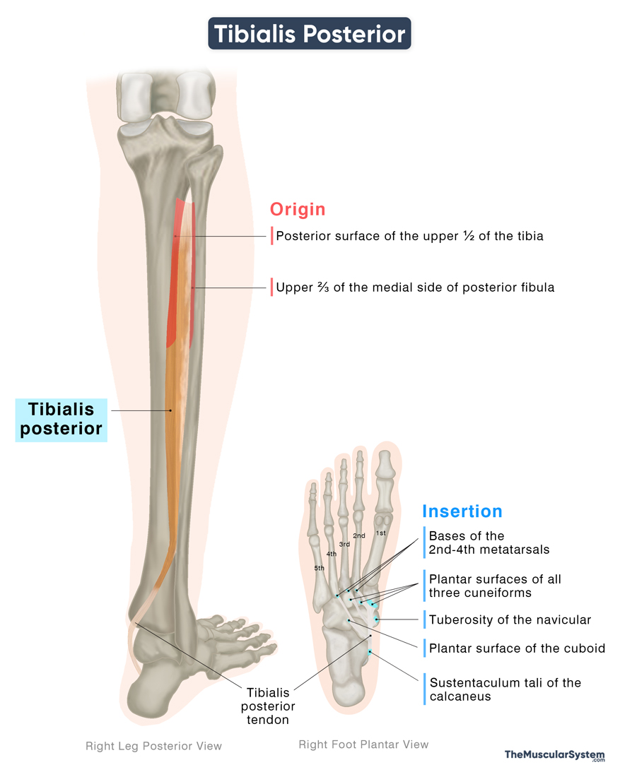
| Origin | — Posterior surface of the upper half of the tibia — Upper two-thirds (2/3) of the medial-posterior fibula — Posterior surface of the interosseous membrane |
| Insertion | — Tuberosity of the navicular bone — Plantar surfaces of all three cuneiforms — Plantar surface of the cuboid bone — Bases of the second to fourth metatarsals — Sustentaculum tali of the calcaneus bone |
Origin
The tibialis posterior muscle has a fleshy belly with a broad origin from both the tibia and fibula. Medially, it arises from the posterior surface of the proximal two-thirds of the tibia, below the soleal line. Laterally, it originates from the posterior surface of the proximal two-thirds of the fibula, particularly along its medial side. In addition, fibers also arise from the posterior surface of the interosseous membrane between the tibia and fibula.
Insertion
From the point of origin, the muscle belly descends medially and narrows into a tendon as it reaches the back of the ankle. Here, the tendon passes through the groove behind the medial malleolus (medial malleolar sulcus), and passes deep to the flexor retinaculum and through the tarsal tunnel. As it exits the tarsal tunnel, the tibialis posterior tendon divides into three components or parts:
- The main part of the tendon attaches chiefly to the tuberosity of the navicular bone, and also to the plantar surface of the medial cuneiform.
- The plantar part spreads across the plantar aspect of the foot to insert into the intermediate and lateral cuneiforms, the cuboid, and the bases of the second to fourth metatarsals.
- The recurrent part, the smallest slip of the tibialis posterior tendon, inserts into the sustentaculum tali, the small bony projection of the medial side of the calcaneus, or heel bone.
Relations With Surrounding Muscles and Structures
Within the deep posterior compartment of the leg, the tibialis posterior is the deepest muscle, occupying a central position between the tibia and fibula. The tibia, fibula, and the interosseous membrane separate it from the muscles in the anterior compartment. The muscle’s belly is covered by the flexor digitorum longus (FDL) on the medial side and the flexor hallucis longus (FHL) on the lateral side. The gastrocnemius, soleus, and plantaris muscles from the superficial posterior compartment are superficial to the tibialis posterior.
At the ankle, as the tendon of tibialis posterior passes behind the medial malleolus, it is the most anterior structure in the tarsal tunnel, followed laterally by the FDL, the posterior tibial artery, the tibial nerve, and the FHL.
Within the sole of the foot, the tendon passes beneath the abductor hallucis and the tendons of flexor digitorum longus. It also gives a slip that contributes to the medial head of flexor hallucis brevis.
Function
| Action | Foot inversion, plantarflexion (ankle), stabilization of medial longitudinal arch |
Its primary action is inversion of the foot at the subtalar and midtarsal joints, turning the sole inward. It works in synergy with the tibialis anterior to perform this movement, which is essential for maintaining balance, particularly when standing or moving on one leg.
The muscle also contributes to plantar flexion at the ankle. Although not the strongest plantar flexor, it assists the gastrocnemius and soleus in pointing the toes downward, such as during walking, running, or rising onto the tiptoes.
Beyond movement, the tibialis posterior plays a crucial structural role. With broad insertions on the navicular, cuneiforms, and metatarsal bones, it supports and elevates the medial longitudinal arch. By reinforcing this arch, the muscle ensures effective weight distribution across the foot and prevents collapse into flatfoot deformity.
Antagonists
Its foot inversion action is antagonized by the fibularis longus and fibularis brevis, which evert the foot. The primary dorsiflexors, the tibialis anterior, extensor hallucis longus, extensor digitorum longus, and fibularis tertius, oppose it in plantar flexion.
Innervation
| Nerve | Tibial nerve (L4-L5) |
The tibialis posterior receives its innervation from the tibial nerve, which branches off the sciatic nerve. This supply comes primarily from the fourth and fifth lumbar nerve roots (L4-L5).
Blood Supply
| Artery | Posterior tibial artery |
Blood supply to the muscle comes from the muscular branches of the posterior tibial artery, a terminal branch of the popliteal artery. Additional supply comes from the fibular (peroneal) artery, a branch of the tibioperoneal trunk.
References
- Anatomy, Bony Pelvis and Lower Limb: Tibialis Posterior Muscle: NCBI.NLM.NIH.gov
- Tibialis Posterior Muscle: Kenhub.com
- Tibialis Posterior Muscle: Radiopaedia.org
- Tibialis Posterior: TeachMeAnatomy.info
- Tibialis Posterior Muscle: Elsevier.com
Della Barnes, an MS Anatomy graduate, blends medical research with accessible writing, simplifying complex anatomy for a better understanding and appreciation of human anatomy.
- Latest Posts by Della Barnes, MS Anatomy
-
Tensor Tympani
- -
Stapedius
- -
Auricularis Posterior
- All Posts


