

Soleus
By
Della Barnes, an MS Anatomy graduate, blends medical research with accessible writing, simplifying complex anatomy for a better understanding and appreciation of human anatomy.
Last updated:
24/09/2025Della Barnes, MS Anatomy
UX/UI Designer at - AdobeDella Barnes, an MS Anatomy graduate, blends medical research with accessible writing, simplifying complex anatomy for a better understanding and appreciation of human anatomy.
What is the Soleus
The soleus is a large, multipennate muscle located at the back of the lower leg, extending from just below the knee to the heel and covering the calf region. Its name comes from “solea,” the Latin word for “sandal.”
Together with the gastrocnemius and plantaris, the soleus forms the superficial posterior compartment of the lower leg. It is also part of the triceps surae, a three-headed muscle group that also includes the gastrocnemius.
Functionally, the soleus is one of the most important muscles of the leg. It plays a key role in supporting the body when standing upright and contributes to walking and other movements.
Anatomy
Location and Attachments
| Origin | On the tibia: The soleal line and the middle 1/3rd of the medial border On the fibula: The head, and the upper 1/4th of the posterior surface of the shaft |
| Insertion | Posterior surface of the calcaneus, via the Achilles tendon |

Origin
The soleus has a complex structure that originates from multiple points on the tibia and fibula. Its primary attachments include:
- The soleal line (also called the popliteal line), an oblique ridge on the upper posterior surface of the tibia.
- The middle one-third of the medial border of the tibia.
- The fibular head and the upper one-fourth of the posterior surface of the fibula.
- The tendinous arch of the soleus, a U-shaped aponeurotic band between the tibia and fibula.
Insertion
The fibers from all the origin points together to form a thick muscle belly that descends toward the ankle, where it joins with the two heads of the gastrocnemius. Together, they give rise to the Achilles tendon, also known as the calcaneal tendon, which is the strongest tendon in the human body. This tendon inserts into the posterior surface of the calcaneus, or the heel bone.
Relations With Surrounding Muscles and Structures
The soleus lies deep to the gastrocnemius, the large muscle forming the visible bulk of the calf. There is an aponeurosis separating the two muscles. When present, the slender and tendinous plantaris muscle also runs between them.
Deep to the soleus is the transverse intermuscular septum, which divides the superficial posterior compartment from the deep posterior compartment of the leg. Beyond this septum, the soleus is related to the deep posterior compartment muscles: tibialis posterior, flexor hallucis longus, and flexor digitorum longus.
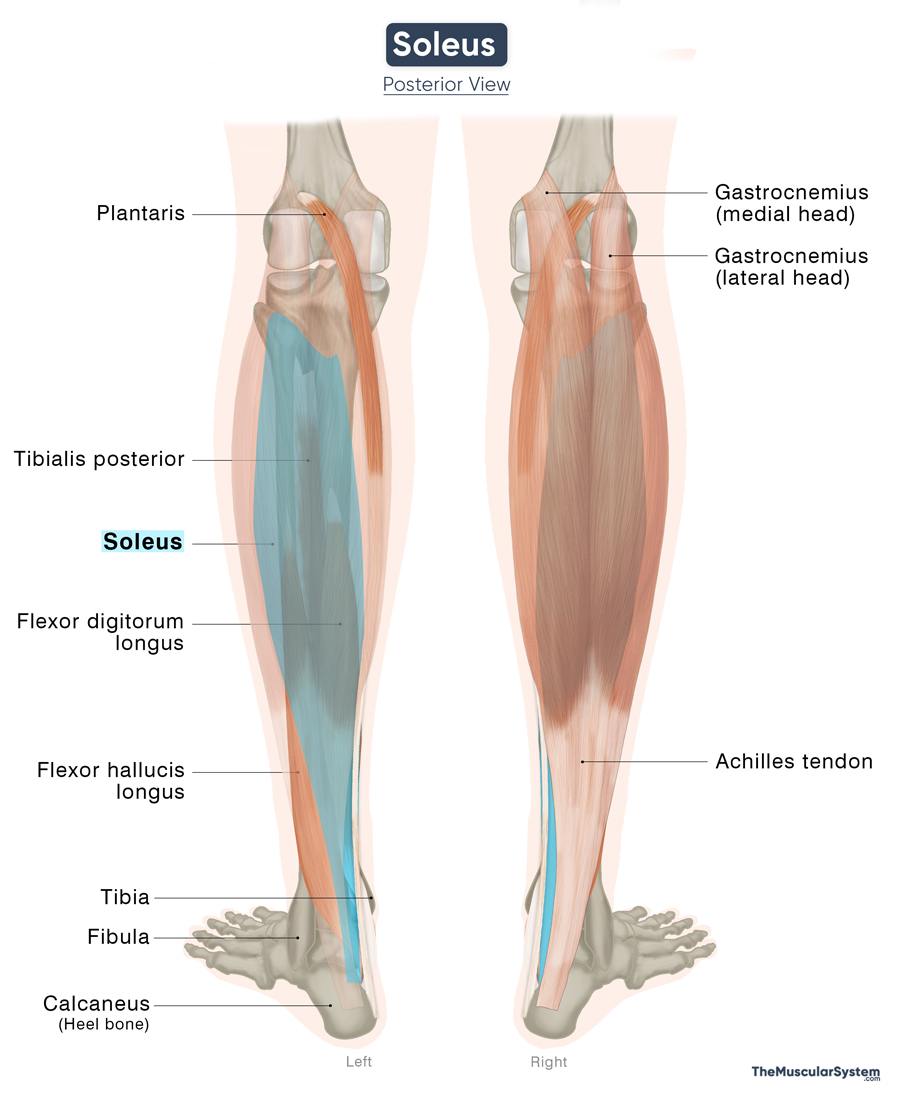
At its proximal attachment, the tendinous arch of the soleus spans between the tibia and fibula. This arch provides passage for the popliteal artery and vein, as well as the tibial nerve, into the posterior compartment of the leg.
Function
| Action | Plantar flexion at the ankle joint |
Plantar Flexion
The soleus and gastrocnemius are the primary muscles to produce plantar flexion at the ankle joint. This movement causes the toes to point downward and allows the heel to lift from the ground, which is essential during walking, running, and jumping. The soleus plays the leading role when the knee is bent, since the gastrocnemius loses much of its effectiveness in that position.
Maintaining Standing Posture
The soleus is often described as an antigravity muscle because of its role in maintaining upright posture. When standing, the body’s center of gravity lies slightly in front of the ankle joint, creating a tendency to fall forward. The soleus counteracts this by maintaining a continuous, low-level contraction that keeps the body balanced. It can effectively perform this task because it is composed mainly of type I (slow-twitch) fibers that resist fatigue and sustain contractions over long periods.
It also makes the muscle important for activities that require stability and fatigue resistance, like walking long distances, hiking, and skiing.
Helping Blood Circulation
The soleus works with the other calf muscles to help push blood back toward the heart. In an upright posture, contraction of the soleus compresses the deep veins of the leg, assisting in propelling blood upward against gravity. For this reason, the calf muscles are sometimes referred to as the “skeletal muscle pump,” or the “peripheral heart,” an action that prevents blood from pooling in the lower limbs and supports circulation during standing and walking.
Antagonists
As the primary dorsiflexor of the ankle joint, the tibialis anterior directly opposes the plantar flexion action of the soleus, making it the principal antagonist of the muscle.
Innervation
| Nerve | Tibial nerve (L5-S2) |
The muscle is innervated by the tibial nerve, which rises from the anterior division of the fifth lumbar to the second sacral nerve roots. The tibial nerve itself is a branch of the sciatic nerve.
Blood Supply
| Artery | Sural, posterior tibial, and peroneal arteries |
The muscle receives its blood supply primarily from two main sources. Proximally, it is supplied by the sural arteries, which arise from the popliteal artery. Distally, it receives branches from both the fibular (peroneal) artery and the posterior tibial artery. The fibular and posterior tibial arteries originate as terminal branches of the popliteal artery within the popliteal fossa.
References
- Soleus Muscle: Kenhub.com
- Soleus Muscle: Radiopaedia.org
- Soleus Muscle: IMAIOS.com
- Soleus: TeachMeAnatomy.info
- Soleus Muscle | Definition, Function & Anatomy: Study.com
- Skeletal Muscle And Respiratory Pumps: IU.Pressbooks.pub
Della Barnes, an MS Anatomy graduate, blends medical research with accessible writing, simplifying complex anatomy for a better understanding and appreciation of human anatomy.
- Latest Posts by Della Barnes, MS Anatomy
-
Tensor Tympani
- -
Stapedius
- -
Auricularis Posterior
- All Posts


