

Popliteus
By
Della Barnes, an MS Anatomy graduate, blends medical research with accessible writing, simplifying complex anatomy for a better understanding and appreciation of human anatomy.
Last updated:
18/09/2025Della Barnes, MS Anatomy
UX/UI Designer at - AdobeDella Barnes, an MS Anatomy graduate, blends medical research with accessible writing, simplifying complex anatomy for a better understanding and appreciation of human anatomy.
What is the Popliteus
The popliteus is a small, thin muscle at the back of the knee joint. It lies in the deep posterior compartment of the lower leg, along with the flexor hallucis longus, flexor digitorum longus, and tibialis posterior. During gait, it plays a key role by unlocking the fully extended knee to initiate flexion. Unlike the other muscles in this compartment, which mainly act on the ankle, the popliteus is the only one that acts on the knee.
Anatomy
Location and Attachments
| Origin | Lateral condyle of the femur |
| Insertion | Posterior surface of the proximal tibia |
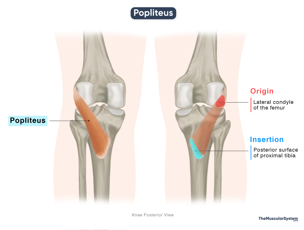
Origin
The popliteus originates from the lateral surface of the lateral femoral condyle through a thick, strong tendon known as the popliteus tendon. Additional fibers may arise from the posterior horn of the lateral meniscus and merge with it, though studies suggest this direct connection is present in only about half of the population.
Insertion
From its origin, the popliteus tendon passes through the popliteal groove and descends medially, expanding into a triangular muscle belly. This belly crosses over the posterior aspect of the knee joint capsule before becoming tendinous again. It inserts along the upper two-thirds of the posteromedial aspect of the proximal tibia. The insertion lies just above the soleal line, where the soleus muscle from the anterior compartment attaches.
Relations With Surrounding Muscles and Structures
The tendon of the muscle is enclosed within the knee joint capsule, which makes it an intracapsular structure. But at the same time, it remains extrasynovial and extra-articular, which means that though it lies inside the capsule, it is outside the synovial cavity and does not contribute to the articular surfaces of the knee.
The popliteus lies deep in the popliteal fossa and forms part of its floor. It is also deep to the fibular collateral ligament, the plantaris, both heads of the gastrocnemius, the tendon of biceps femoris, and the neurovascular bundle composed of the popliteal vessels and tibial nerve.
In addition, the popliteus has an indirect connection to the fibula through the popliteofibular ligament, which extends from its tendon to the fibular head.
Variation
- Sometimes, the popliteus has an extra head arising from the sesamoid bone in the lateral head of the gastrocnemius.
- A small sesamoid bone, the cyamella, can occasionally be found within the popliteus tendon. Studies on this bone are limited, but available evidence suggests it occurs in less than 2% of the population.
- Sometimes, a small accessory muscle, the popliteus minor, arises from the medial side of the plantaris muscle on the distal femur and inserts into the posterior ligament of the knee joint.
- The peroneal tibialis is another minor accessory muscle that may lie beneath the popliteus, starting from the fibular head and extending to the soleal line on the tibia.
Function
| Action | Unlocks the knee by rotating the tibia/femur to assist flexion of the fully extended knee joint. |
The popliteus muscle is most important at the beginning of knee flexion. While it does not produce much flexion force, it is essential for unlocking the fully extended knee. For this reason, it is often called the “key” for unlocking the knee.
In the closed-chain phase of the gait cycle, when the foot is fixed on the ground, the tibia is slightly rotated laterally on the femur, which locks the joint for stability. To begin flexion, the popliteus contracts and rotates the femur laterally (outward) on the fixed tibia, unlocking the knee. This action allows the knee to bend smoothly from its stable, locked position.
In the open-chain phase, when the foot is free to move, the popliteus acts on the tibia instead, rotating it medially (inward) on the femur. This aligns the tibia with the femoral condyles and stabilizes the joint surfaces, making flexion smooth and controlled during the swing phase of gait.
Beyond initiating motion, the popliteus also stabilizes the posterior knee through its connections to the joint capsule and supporting ligaments. It further protects the lateral meniscus by drawing it backward during flexion, preventing it from being trapped between the femur and tibia.
Antagonists
The primary antagonists to the popliteus muscle are the quadriceps muscles (rectus femoris, vastus lateralis, vastus medialis, vastus intermedius), which extend the knee, and the biceps femoris, which laterally rotates the tibia in opposition to the popliteus.
Innervation
| Nerve | Tibial nerve (L4-S1) |
The muscle is innervated by the tibial nerve, which rises from the fourth and fifth lumbar and the first sacral nerve roots. It is a branch of the sciatic nerve.
Blood Supply
| Artery | Inferior medial and inferior lateral genicular arteries |
The muscle receives its main blood supply from the inferior medial and inferior lateral genicular arteries, which are branches of the popliteal artery. Additional contributions may come from the posterior tibial recurrent artery, the proximal portion of the posterior tibial artery, and occasionally from the nutrient artery of the tibia.
References
- Popliteus Muscle | Action, Function & Location: Study.com
- Popliteus Muscle: Kenhub.com
- Anatomy, Bony Pelvis and Lower Limb: Popliteus Muscle: NCBI.NLM.NIH.gov
- Popliteus: TeachMeAnatomy.info
- Popliteus: Rad.UW.edu
- Popliteus Muscle: IMAIOS.com
Della Barnes, an MS Anatomy graduate, blends medical research with accessible writing, simplifying complex anatomy for a better understanding and appreciation of human anatomy.
- Latest Posts by Della Barnes, MS Anatomy
-
Tensor Tympani
- -
Stapedius
- -
Auricularis Posterior
- All Posts


