

Extensor Digitorum
By
Della Barnes, an MS Anatomy graduate, blends medical research with accessible writing, simplifying complex anatomy for a better understanding and appreciation of human anatomy.
Last updated:
05/05/2023Della Barnes, MS Anatomy
UX/UI Designer at - AdobeDella Barnes, an MS Anatomy graduate, blends medical research with accessible writing, simplifying complex anatomy for a better understanding and appreciation of human anatomy.
What is Extensor Digitorum
The Extensor digitorum also referred to as extensor digitorum communis (EDC), is one of the 6 superficial extensor muscles in the posterior compartment of the forearm. The other 5 are the extensor digiti minimi, extensor carpi ulnaris, brachioradialis, and the extensor carpi radialis longus and brevis.
It is a long muscle coursing from the forearm to the hand and fingers, thus being considered an extrinsic muscle of the hand.
Anatomy
Location and Attachments
| Origin | Lateral epicondyle of the humerus (the common extensor tendon) |
| Insertion | The extensor expansion of the 2nd to 5th digits |
Origin
Like most other superficial posterior muscles, the extensor digitorum originates from the common extensor tendon at the lateral epicondyle of the humerus. After originating, the muscle courses distally along the back of the forearm. Once the muscle belly reaches the distal 1/3 of the forearm, just above the wrist region, it divides into 4 tendons. These tendons then travel further down the forearm to enter the back surface of the hand (dorsum) and pass deep to the extensor retinaculum. The extensor retinaculum is a thick fibrous band of ligaments at the back of the wrist that holds the tendons of the hand’s extensors together.
Insertion
After passing through the extensor retinaculum, the 4 tendons course towards their corresponding digits, from 2nd to 5th, and spread out to insert into the extensor expansions. Also known as the extensor hoods, these are the triangular aponeuroses that cover the back of the 2nd to 5th digits, between the metacarpophalangeal and proximal interphalangeal joints. The extensor expansions are also joined by the tendons of the other primary digital extensors, the lumbricals, and interossei.
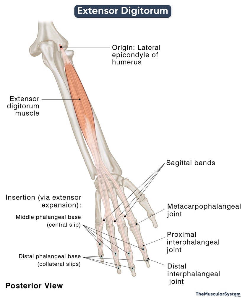
At the proximal interphalangeal joint, each extensor expansion divides into 3 slips with 1 central and 2 collateral parts. The central part goes on to insert into the back surface of the middle phalangeal base of each digit. The collateral parts continue a little further along the sides of the digits to insert into the base of the distal phalanges on the posterior surface.
The tendons to the 3rd, 4th, and 5th digits (middle, ring, and little fingers) are linked with each other on the dorsal side of the hand by 2 slanting or oblique intertendinous bands. These fibrous connective bands, known as sagittal bands, may sometimes show anatomical variations.
Relations With Surrounding Muscles and Structures
It is the most superficial of all the posterior forearm muscles, with the deep posterior muscles, abductor and extensor pollicis longus lying immediately deep to it. The space enclosed by these 3 muscles allows passage to the posterior interosseous artery and nerve.
In the forearm, it has the extensor carpi radialis brevis on its lateral side, while the extensor digiti minimi and extensor carpi ulnaris are located on its medial side.
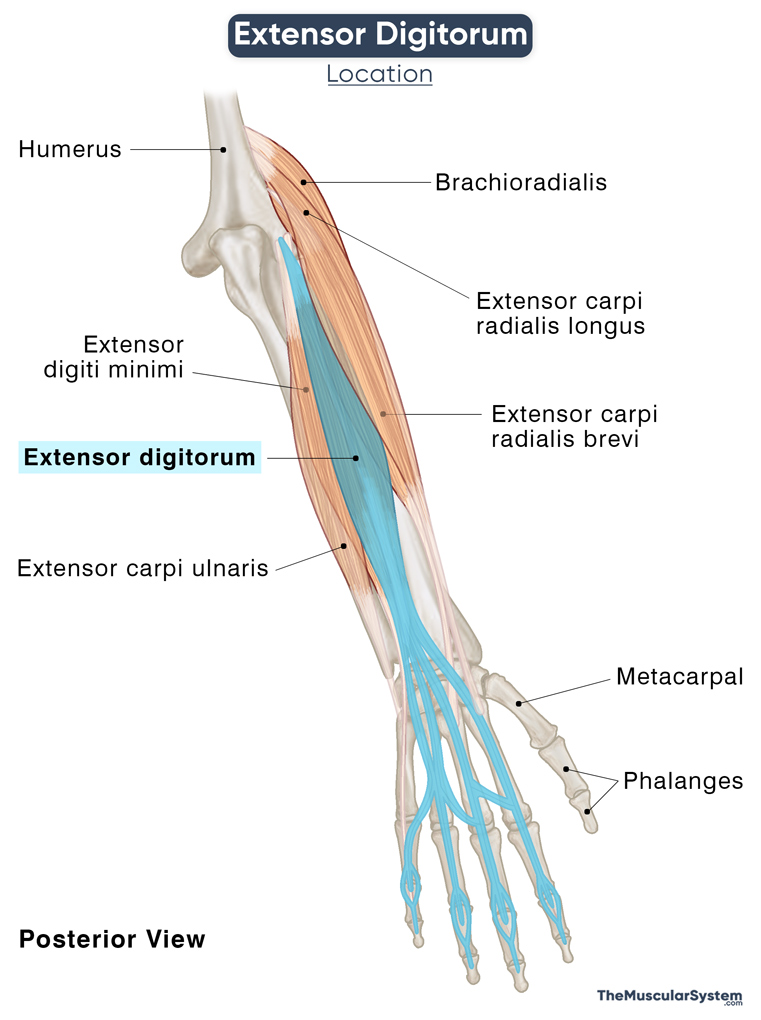
The long inserting tendon lies superficial to the dorsal interossei muscles. It runs deep to the extensor retinaculum, coursing between the extensor digiti minimi and extensor pollicis longus tendons. The extensor digitorum tendons, accompanied by the extensor indicis, fill the 4th extensor compartment on the dorsal surface of the hand.
Function
| Action | Extension of the fingers at all the joints |
- As evident from its name, the extensor digitorum’s main action is to extend the index, middle, ring, and little fingers at the metacarpophalangeal and the two interphalangeal joints.
- As well as extending the fingers, the muscle helps spread each of the 4 finders out when the palm is open.
- The muscle is active when you grip something in your hand because its contraction allows you to open the hand and let go.
- It also plays a minor role in extending the wrist joint.
The extensor digitorum works against two antagonists, the forearm flexors, flexor digitorum profundus, and flexor digitorum superficialis.
Innervation
| Nerve | Posterior interosseous nerve (C7 and C8) |
The posterior interosseous nerve (C7, C8) that continues from the radial nerve’s deep branch innervates the extensor digitorum.
Blood Supply
| Artery | Posterior interosseous, anterior interosseous, and radial recurrent arteries |
Blood supply to the muscle comes from 3 different sources, the anterior and posterior interosseous arteries and the radial recurrent artery. The first two are branches of the ulnar artery, while the third rises from the radial artery.
References
- Extensor Digitorum Muscle: GetBodySmart.com
- Extensor Digitorum: IMAIOS.com
- Extensor Digitorum Muscle: KenHub.com
- Extensor Digitorum Muscle: RadioPaedia.org
- Extensor Digitorum: HealthLine.com
Della Barnes, an MS Anatomy graduate, blends medical research with accessible writing, simplifying complex anatomy for a better understanding and appreciation of human anatomy.
- Latest Posts by Della Barnes, MS Anatomy
-
Tensor Tympani
- -
Stapedius
- -
Auricularis Posterior
- All Posts


