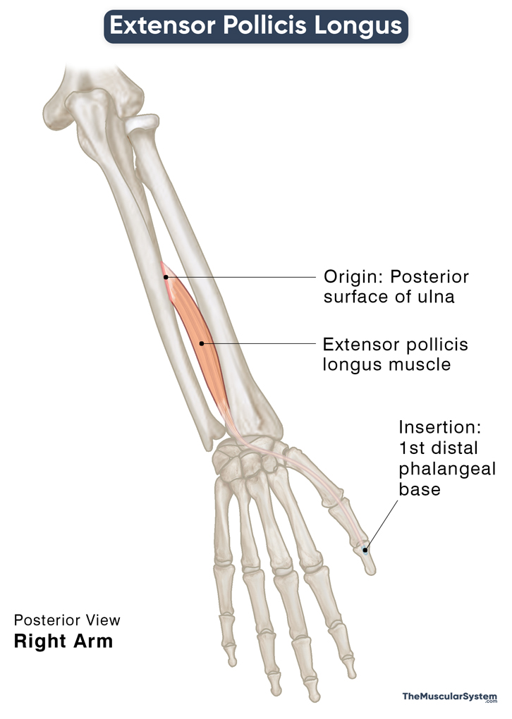
Extensor Pollicis Longus
Last updated:
05/06/2024Della Barnes, an MS Anatomy graduate, blends medical research with accessible writing, simplifying complex anatomy for a better understanding and appreciation of human anatomy.
What is Extensor Pollicis Longus
The extensor pollicis longus (EPL) is one of the 5 deep forearm muscles in the posterior compartment, along with the supinator, extensor indicis, abductor pollicis longus, and extensor pollicis brevis. The extensor pollicis longus is an extrinsic muscle of the thumb and one of the muscles enclosing the anatomical snuff box (the triangular depression at the thumb’s base).
Anatomy
Location and Attachments
| Origin | The posterior surface of the middle of ulna, and the interosseus membrane |
| Insertion | The posterior surface of the base of the 1st distal phalanx |
Origin
The long, slender muscle originates from the middle one-third of the ulna, along its posterior surface and radial border. The point of origin extends to the interosseus membrane, the band of connective tissue that obliquely runs between the ulna and radius. The origin of the EPL lies proximal to that of the extensor indicis muscle.
After originating, the muscle belly runs distally, coursing obliquely towards the radial side of the wrist.
Insertion
As it passes the wrist joint, the muscle belly narrows into a long tendon. This EPL tendon passes over the Lister’s tubercle before traveling underneath the extensor retinaculum. The Lister’s tubercle, the bony prominence on the dorsal surface of the radius’s distal end, works as a pulley as the tendon passes over it and then pivots to course obliquely in the thumb’s direction.
The EPL tendon solely occupies the 3rd extensor compartment of the wrist.
Once the tendon enters the dorsum of the hand, it travels further distally and laterally to insert into the posterior surface of the thumb’s distal phalangeal base.
Relations With Surrounding Muscles and Structures
It is a deep muscle in the posterior forearm, with the extensor digitorum lying superficial at its proximal end. The abductor pollicis longus lies on the lateral side of the EPL, while the extensor indicis lies on the medial side.
On the side of the wrist joint, its long tendon crosses the tendons of the superficial forearm muscles, extensor carpi radialis longus, and brevis at the back of the hand.
The extensor pollicis longus is the third muscle in the anatomical snuff box, forming its medial border. The extensor pollicis brevis, the muscle on its lateral side, forms the radial border along with the abductor pollicis longus.
Before its insertion, it comes together with the tendons of the adductor pollicis (medially) and the abductor pollicis brevis (laterally). These 3 tendons contribute to the extensor expansion of the thumb, which takes care of all extending movements of the thumb at all its joints.
The posterior interosseous nerve and artery run across the back of the EPL muscle.
Function
| Action | Extension of the thumb at all its joints |
As clearly suggested by its name, it is an extensor of the thumb. Its primary function is to help extend the thumb at the metacarpophalangeal (MCP) joint and its single interphalangeal (IP) joint. It synergizes with the extensor pollicis brevis when extending the MCP joint.
Once the thumb is fully extended, the EPL also plays a role in adducting the metacarpal of the thumb. All these movements are crucial to the functioning of the thumb and, thus, the hand.
It also has some minor functions for moving the wrist and forearm. With its tendon crossing the distal radioulnar joint (wrist), the extensor pollicis longus help extend and abduct the wrist along with the other thumb extensors. It also assists in supinating the forearm or rotating it to face upward.
Innervation
| Nerve | Posterior interosseous nerve (C7 and C8) |
The posterior interosseous nerve (C7, C8) that innervates this muscle is a branch of the radial nerve via its deep branch.
Blood Supply
| Artery | Posterior and anterior interosseous arteries |
The primary blood supply to the muscles comes from the posterior interosseous artery, while the anterior interosseous artery (perforating branches) offers additional support. Both of these arteries are branches of the ulnar artery.
References
- Anatomy, Shoulder and Upper Limb, Hand Extensor Pollicis Longus Muscle: NCBI.NLM.NIH.gov
- Extensor Pollicis Longus Muscle: KenHub.com
- Extensor Pollicis Longus: IMAIOS.com
- Extensor Pollicis Longus: TeachMeAnatomy.info
- Extensor Pollicis Longus Muscle: RadioPaedia.org
Della Barnes, an MS Anatomy graduate, blends medical research with accessible writing, simplifying complex anatomy for a better understanding and appreciation of human anatomy.
- Latest Posts by Della Barnes, MS Anatomy
-
Infrahyoid Muscles
- -
Omohyoid
- -
Sternohyoid
- All Posts


