

Abductor Pollicis Longus
By
Della Barnes, an MS Anatomy graduate, blends medical research with accessible writing, simplifying complex anatomy for a better understanding and appreciation of human anatomy.
Last updated:
05/06/2024Della Barnes, MS Anatomy
UX/UI Designer at - AdobeDella Barnes, an MS Anatomy graduate, blends medical research with accessible writing, simplifying complex anatomy for a better understanding and appreciation of human anatomy.
What is the Abductor Pollicis Longus
The abductor pollicis longus, or APL, is a muscle in the deep posterior compartment of the forearm. It is one of the three muscles that form the anatomical snuff box. The other two muscles in this group are the extensor pollicis brevis and longus. It is an extrinsic muscle of the hand.
Anatomy
Location and Attachments
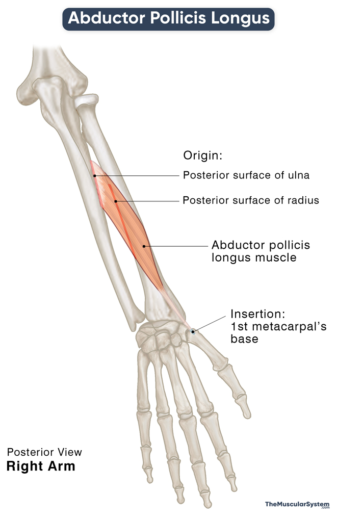
| Origin | The posterior surfaces of the ulna and radius, along with the interosseus membrane between them |
| Insertion | The base of the 1st metacarpal (thumb) |
Origin
The elongated muscle has a relatively broad point of origin, spanning the surfaces of both the ulna and radius. On the ulna, it originates from the proximal third of the dorsal surface. On the radius, it arises from the middle third of the dorsal surface. And since these two bones are connected via the oblique fibers of the interosseus membrane, some part of the muscle’s origin also extends to this membrane. The origin of the APL lies just below the insertion point of the anconeus, the upper arm muscle.
Insertion
The muscle belly runs distally and laterally towards the radial side, becoming more superficial as it reaches the distal end of the forearm. It narrows into a tendon before it reaches the wrist, which then continues distally towards the hand.
Near the wrist joint, the APL tendon passes through the groove at the distal end of the radius on the lateral surface. Here, it is joined by the tendon of the extensor pollicis brevis. The two tendons continue further down to share the 1st extensor compartment as they pass underneath the extensor retinaculum to enter the hand on its dorsal side.
At this point, the tendon gives off multiple branches or slips that insert at different points — the main part inserts into the base of the 1st metacarpal (the thumb’s metacarpal). The slips insert into the surrounding structures, including the trapezium bone, the opponens pollicis, and abductor pollicis brevis muscles in the hand, and the fascia of the thenar eminence (the fleshy mound at the base of the thumb). The number of these slips often varies between individuals.
Relations With Surrounding Muscles and Structures
The belly of the abductor pollicis longus has the extensor digitorum lying superficial to it. The extensor pollicis longus lies medial to the APL belly, while the extensor pollicis brevis tendon lies medial to the APL tendon.
As the muscle travels distally down the forearm and passes the extensor retinaculum, it gradually becomes more superficial. Its tendon, along with the extensor pollicis brevis, forms the lateral border (the radial side) of the anatomical snuffbox. The anatomical snuffbox is the triangular depression that becomes evident at the base of the thumb in the back of your hand when you extend or abduct the thumb. It allows passage to the neurovascular bundles of the hand.
The abductor pollicis longus and the extensor digitorum allow the posterior interosseous artery and nerve to pass between them and travel over the superficial surface of the APL.
Function
| Action | Extension and abduction of the thumb at the carpometacarpal joint |
Abducting the Thumb at the Carpometacarpal Joint
The primary function of the abductor pollicis longus is abducting the thumb at the 1st carpometacarpal joint, which means moving the thumb in any direction away from the rest of the hand. The abductor pollicis brevis helps the APL in this action.
An example of this movement is the way you hold a bowling ball before launching it.
Extending the Thumb at the Metacarpophalangeal Joint
It works alongside the extensor pollicis longus and brevis to extend the metacarpophalangeal joint, which fully extends the thumb. This action helps you let go of an object you are holding in your hand.
Extending and Abducting the Wrist
An additional function of the muscle is to help the wrist abductors with radial deviation of the wrist at the radiocarpal joint. It means bending the wrist to the thumb’s side, a movement essential for multiple actions of the forearm and hand.
The primary antagonist of the abductor pollicis longus is the adductor pollicis, a muscle in the hand.
Innervation
| Nerve | Posterior interosseous nerve (C7 and C8) |
The posterior interosseous nerve, with the nerve roots C7 and C8, arises from the radial nerve via its deep branch.
Blood Supply
| Artery | Posterior interosseous arteries |
The proximal part of the muscle receives vasculature from the posterior interosseous artery’s lateral branch. An additional smaller supply comes to the distal part of the muscle from the anterior interosseous artery via its perforating branch. Both these arteries branch off from the ulnar artery.
References
- Abductor Pollicis Longus Muscle: KenHub.com
- Abductor Pollicis Longus: Origin, Insertion, & Innervation: Study.com
- Abductor Pollicis Longus: TeachMeAnatomy.info
- Abductor Pollicis Longus: IMAIOS.com
- Abductor Pollicis Longus Muscle: RadioPaedia.org
- Abductor Pollicis Longus: Rad.Washington.edu
- Abductor Pollicis Longus: EatonHand.com
Della Barnes, an MS Anatomy graduate, blends medical research with accessible writing, simplifying complex anatomy for a better understanding and appreciation of human anatomy.
- Latest Posts by Della Barnes, MS Anatomy
-
Tensor Tympani
- -
Stapedius
- -
Auricularis Posterior
- All Posts


