

Supinator
By
Della Barnes, an MS Anatomy graduate, blends medical research with accessible writing, simplifying complex anatomy for a better understanding and appreciation of human anatomy.
Last updated:
23/07/2026Della Barnes, MS Anatomy
UX/UI Designer at - AdobeDella Barnes, an MS Anatomy graduate, blends medical research with accessible writing, simplifying complex anatomy for a better understanding and appreciation of human anatomy.
What is Supinator
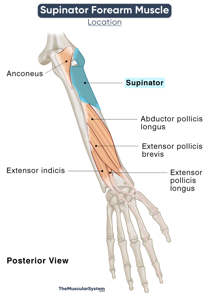
The supinator is a broad, spiral muscle in the deep posterior compartment of the forearm. It is one of the 5 muscles in this compartment, with the others being the extensor indicis, abductor pollicis longus, and the extensor pollicis longus and brevis.
Anatomy
Location and Attachments
| Origin | Lateral epicondyle of humerus, supinator crest of ulna, radial collateral ligament, and annular ligament |
| Insertion | The posterior surface of radius |
The supinator is curved or wrapped around the proximal third of the radius bone. It has points of origin on all the arm bones, humerus, radius, and ulna.
Origin
The muscle has two layers or heads, superficial and deep, originating from the same points. The difference between the two is that the superficial layers originate via tendons, while the deep layer arises via muscular slips.
- On the humerus, the muscle arises from the lateral epicondyle.
- On the ulna, it attaches to the supinator crest, the oblique ridge running down the posterior surface of the bone from the radial notch. A few of its fibers also originate from the adjoining ulnar fossa.
- On the radius, the two layers arise from the radial tuberosity via the radial collateral ligament and the annular ligament. The superficial layer wraps around the proximal end of the bone, while the upper part of the deep layer surrounds its neck like a sling.
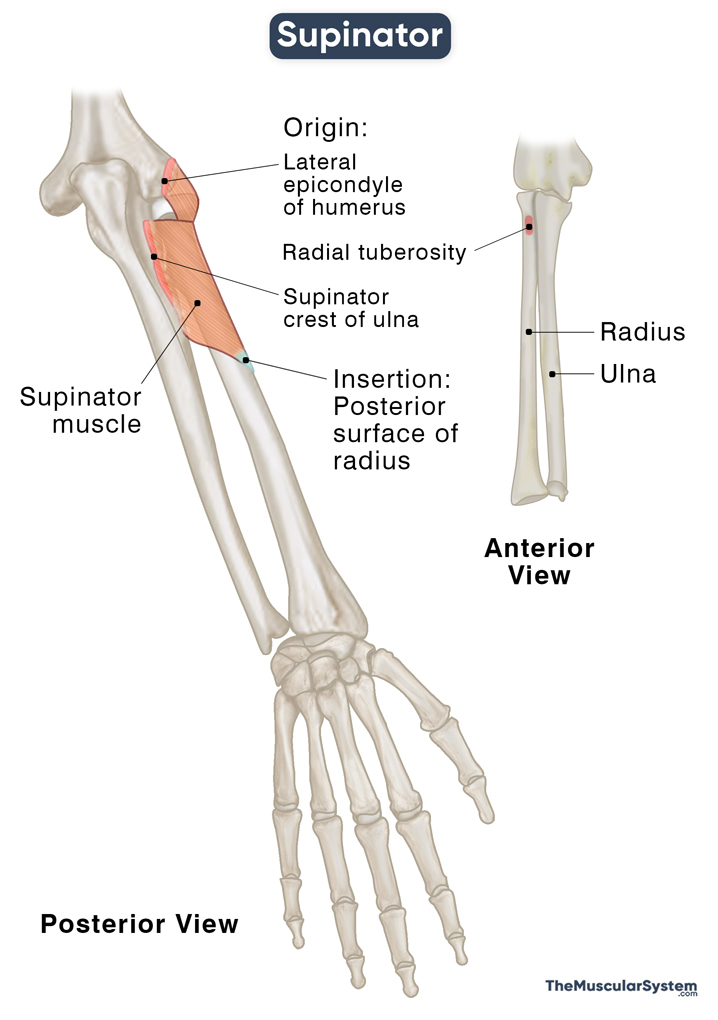
Insertion
The superficial layer of the muscle inserts into that radius at the lateral border of the radial tuberosity and the oblique line. This attachment lies almost at the same point as the insertion of the superficial forearm flexor pronator teres.
The deep layer inserts into the proximal third of the radius at the back of its medial surface, between the head of radius and the oblique line.
Relations With Surrounding Muscles and Structures
As mentioned in the beginning, the supinator is a deep muscle lying directly over the radius and ulna where it attaches.
It forms the distal part of the floor of the cubital fossa, the triangular depression inside the elbow joint. The upper arm’s brachialis muscle forms the floor’s proximal part.
The lower end of the anconeus muscle lies superficial to the supinator on its ulnar (little finger) side. The superficial muscles in the posterior forearm, extensor carpi radialis longus, and brevis lay over its radial (thumb-side) surface.
The superficial and deep layers or heads have an opening between them to pass the radial nerve’s deep branch just before it enters the posterior compartment of the forearm.
Sometimes there is a band of ligament, known as the oblique cord, crossing over the muscle’s deep head. This ligament has no apparent use, but when present, it connects the radius and ulna at their tuberosities.
Function
| Action | Supination of the forearm |
As the name ‘supinator’ clearly suggests, it is the primary supinator of the forearm.
The muscle originates near the elbow joint and wraps around the radius before attaching to multiple points on its surface. It means when the supinator contracts, it pulls the radius to rotate it at the proximal radioulnar joint. This action of the muscle on the radius results in the supination of the forearm, which means rotating it so the palm faces upward.
The action of the supinator is enough to supinate the forearm for regular activities like holding a pen to write, carrying a tray of food, or holding your phone to your ear.
When you need to supinate the forearm quickly and forcefully or against resistance, the biceps brachii assists the supinator. However, the biceps cannot supinate the forearm when fully extended. It is most effective when the elbow is already flexed by at least 90°, such as when using a screwdriver.
Innervation
| Nerve | Posterior interosseous nerve (C7 and C8) |
The posterior interosseous nerve, which is the deep branch of the radial nerve, arising from nerve roots C7 and C8, supplies this muscle. Nerve branches arising from nerve roots C5 and C6 may also be involved in the muscle’s innervation.
Blood Supply
| Artery | Radial recurrent artery, posterior interosseous artery, posterior interosseous recurrent arteries |
The superior layer or head of the muscle is supplied by the radial recurrent branch of the radial artery.
The deep layer receives vasculature from the posterior interosseous artery, and the posterior interosseous recurrent artery — both branches of the ulnar artery.
References
- Supinator: IMAIOS.com
- Supinator Muscle: RadioPaedia.org
- Supinator Muscle: KenHub.com
- Supinator Function and Anatomy: Study.com
- Supinator: TeachMeAnatomy.info
- Supinator: Rad.Washington.edu
Della Barnes, an MS Anatomy graduate, blends medical research with accessible writing, simplifying complex anatomy for a better understanding and appreciation of human anatomy.
- Latest Posts by Della Barnes, MS Anatomy
-
Tensor Tympani
- -
Stapedius
- -
Auricularis Posterior
- All Posts


