Pronator Teres
Last updated:
28/05/2024Della Barnes, an MS Anatomy graduate, blends medical research with accessible writing, simplifying complex anatomy for a better understanding and appreciation of human anatomy.
What is the Pronator Teres
The pronator teres is one of the five superficial flexor muscles in the anterior compartment of the forearm. Its name is derived from the Latin word ‘pronator,’ which means ‘lying face down.’ It refers to the main function of the muscle, which is rotating the forearm for the palm to face down.
Anatomy
Location and Attachments
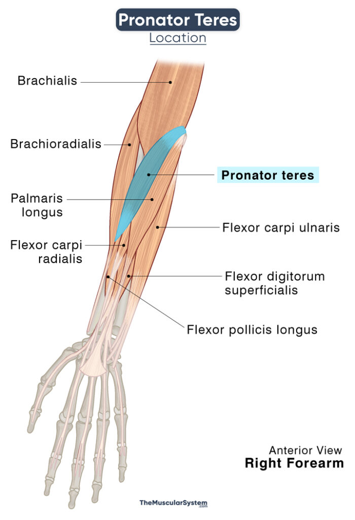
This fusiform muscle in the forearm’s anterior compartment is the most lateral of the superficial flexor muscles, the other four in the group being the flexor digitorum superficialis, palmaris longus, flexor carpi radialis, and flexor carpi ulnaris.
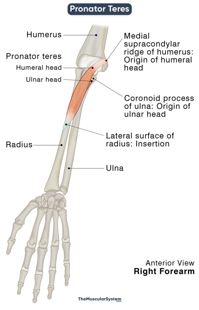
Pronator teres has two heads, the humeral and the ulnar, named after their origin.
| Origin | Humeral head: Medial supracondylar ridge of the humerus Ulnar head: Coronoid process of the ulna |
| Insertion | The middle part of the lateral surface of radius |
The humeral head is the larger of the two, lying superficial to the ulnar head. As the name suggests, it originates from the humerus’s medial supracondylar ridge above the medial epicondyle, as well as via the common flexor tendon. The ulnar head originates from the ulna’s coronoid process.
The two heads come together to form the muscle belly, which courses distally to insert into the lateral surface of the radius, on the pronator tuberosity, to be more specific.
Relations With Surrounding Muscles and Structures
The muscle belly of the pronator teres lies deep the brachioradialis muscle in the anterior compartment’s superficial layer, while its proximal part is underneath the flexor digitorum superficialis. The muscle is located laterally from and directly beside the flexor carpi radialis muscle.
The point of origin of the ulnar head lies between the insertion point of the brachialis and the origin of the radial head of flexor digitorum superficialis, just above where the flexor pollicis longus originates.
The pronator teres constitutes the medial margin of one of the arm’s critical anatomical landmarks, the cubital fossa or elbow pit. As is evident from the name, it is the small triangular area on the inner side of the elbow, between the upper and lower arms. This region holds multiple neurovascular structures related to the pronator teres.
The radial and ulnar arteries branch out from the brachial artery near the superior margin of pronator teres. Afterward, the radial artery runs along the muscle’s superior edge, while the ulnar artery courses from its posterior side. The medial nerve passes between the humeral and ulnar heads, with the latter separating it from the ulnar artery.
Function
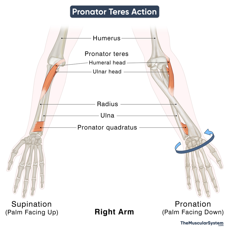
| Action | Pronation of the forearm, flexion of the elbow joint (minor) |
- Pronation of the Forearm: It is the primary function of the muscle, the one it is named after. The muscle acts as a synergist to the pronator quadratus and medially pulls the radius, causing the head of the bone to rotate around the ulna at the proximal radioulnar joint. The movement causes the forearm to rotate inwards, bringing the palm to a pronated position, facing the ground. Due to this action, the pronator teres is vital in various sporting activities.
- Flexion at the Elbow: Since the muscle has synergistic effects with the biceps brachii, brachialis, and brachioradialis, the primary flexors of the forearm at the elbow, the pronator teres itself lends some assistance with the movement.
So, the muscle is essential for small everyday activities like eating, writing, and rotating your hand to put something down or pick something up.
The primary antagonist of pronator teres is the supinator muscle. The biceps brachii also works as an antagonist to pronation.
Innervation
| Nerve | Median nerve (C6 and C7), a branch of the brachial plexus (C5-T1) |
Blood Supply
| Artery | Branches of the ulnar, radial, and brachial arteries |
The branches of the ulnar artery that supply to the pronator teres are the anterior ulnar recurrent artery and common interosseus artery. The muscle also receives blood supply from the radial artery branch, radial recurrent artery, and brachial artery branches, inferior ulnar collateral arteries.
References
- Pronator Teres: Definition, Function & Nerve Supply: Study.com
- Anatomy, Shoulder and Upper Limb, Pronator Teres: NCBI.NLM.NIH.gov
- Pronator Teres – Attachments, Action & Innervation: GetBodySmart.com
- Pronator Teres: Rad.Washington.edu
- Pronator Teres Muscle: KenHub.com
- Pronator Teres: Meddean.LUC.edu
Della Barnes, an MS Anatomy graduate, blends medical research with accessible writing, simplifying complex anatomy for a better understanding and appreciation of human anatomy.
- Latest Posts by Della Barnes, MS Anatomy
-
Extensor Digitorum Brevis
- -
Extensor Hallucis Brevis
- -
Posterior Compartment of the Leg
- All Posts


