

Palmaris Longus
By
Della Barnes, an MS Anatomy graduate, blends medical research with accessible writing, simplifying complex anatomy for a better understanding and appreciation of human anatomy.
Last updated:
23/07/2026Della Barnes, MS Anatomy
UX/UI Designer at - AdobeDella Barnes, an MS Anatomy graduate, blends medical research with accessible writing, simplifying complex anatomy for a better understanding and appreciation of human anatomy.
What is the Palmaris Longus
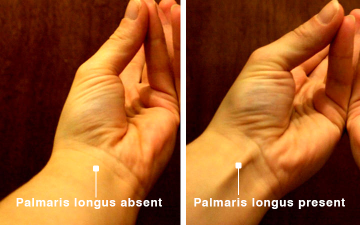
The palmaris longus, or PL, is a long, spindle-shaped, superficial, extrinsic muscle in the anterior compartment of the forearm. The muscle may be absent altogether in 3-15% of people, with the numbers varying based on population and ethnicity. When present, it weakly acts on wrist flexion. It is one of the five superficial flexors of the forearm, along with pronator teres, flexor carpi radialis, flexor carpi ulnaris, and flexor digitorum superficialis.
Anatomy
Location and Attachment
| Origin | Medial epicondyle of the humerus (the common flexor tendon) |
| Insertion | Distal side of the flexor retinaculum, and the palmar aponeurosis |
Like the other superficial muscles of the anterior compartment, the palmaris longus originates from the common flexor tendon at the medial epicondyle. The origin tendons broaden into a small muscle belly, which flattens into a slender tendon around the middle third of the forearm.
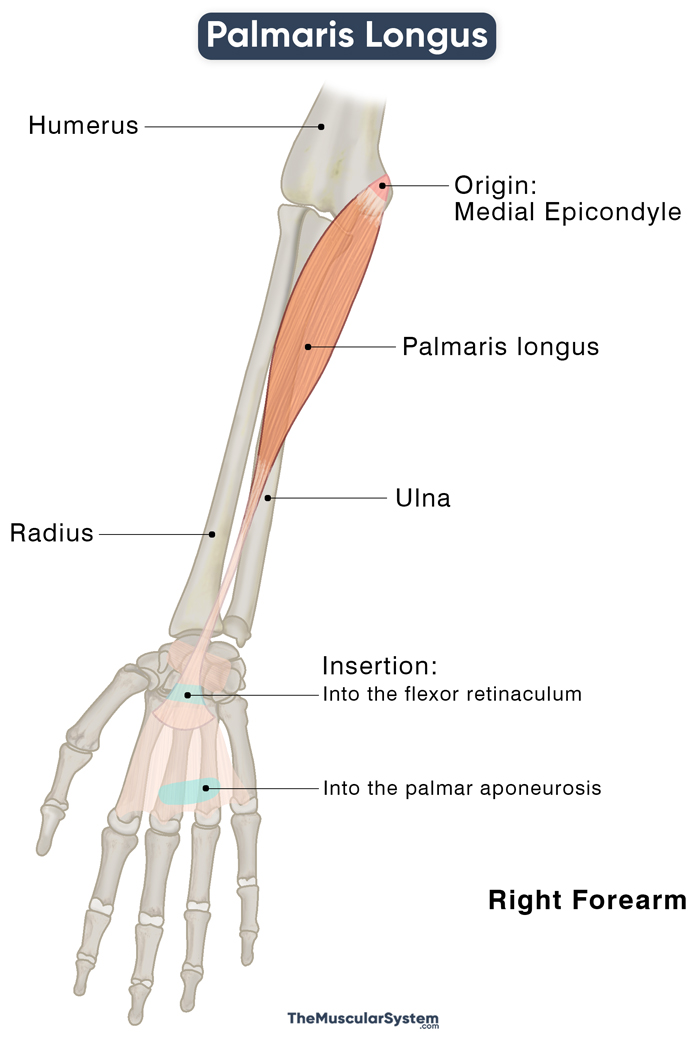
This long tendon courses down to enter the surface of the palm, and passes the flexor retinaculum (the fibrous band of connective tissue that forms carpal tunnel’s anterior roof) where a few of its fibers fuse. The rest of the tendon continues distally and inserts on the palmar aponeurosis part of the palm’s deep fascia.
Anatomical Variation
It often shows different anatomic variations, the most common being the absence of the muscle in one (unilateral) or both (bilateral) arms. As mentioned above, studies show ethnicity as a possible factor in this, as the muscle has been found to be absent in around 15% of Caucasians and Hispanics, around 5% of African-Americans, and around 2.5-3% of Asians. Gender might also be a factor, as studies show it to be more commonly absent in females than in males, at least in some ethnic groups.
The muscle may also have differences in the length of the tendons and muscle belly, and sometimes the whole muscle may present just as a long tendon. Variation in the point of insertion, like the antebrachial fascia, the tendons of the flexor carpi ulnaris that insert into the pisiform bone, and the navicular bone, may also be evident. The reason for these variations needs to be clarified.
Its absence shows no effect on the grip and functioning of the wrist and hand. However, those with the muscle missing have reduced pinch strength in the 4th and 5th fingers.
Relations With Surrounding Muscles and Structures
Palmaris longus lies deep only to the layer of the skin. The flexor digitorum superficialis lies deep to the muscle. The flexor carpi radialis and the flexor carpi ulnaris are located on the two sides of the palmaris longus.
At the wrist, the muscle lies superficial to the median nerve. The flexor carpi radialis tendon runs medial to the tendon of the palmaris longus, allowing the median nerve passage between the two.
Functions
| Action | Flexion of the wrist (weak), tightening the palmar aponeurosis |
Flexing the Wrist: Since the muscle runs the length of the forearm, from the medial epicondyle to the wrist, anatomically, it should contribute to flexing the wrist joint. However, the small and narrow muscle can only assist the flexor carpi radialis and flexor carpi ulnaris, being an accessory flexor of the wrist itself.
Tightening the Grip: When the muscle contracts, the tendon fibers that attach to the palmar aponeurosis tighten this connective tissue sheath which causes the 2nd to 5th metacarpophalangeal joints to flex slightly. This action pulls the sheath, and the skin towards the wrist, ensuring that the skin layer remains tightly in place instead of coming off like a glove when the wrist moves. It also helps grip something in your hand.
Though not critical to the functioning of hands, the muscle has considerable clinical significance as it is often used to repair other tendons in the upper limb (autograft).
Innervation
| Nerve | Median nerve (C7 and C8) |
The median nerve (C7 and C8) branch rising from the brachial plexus’s medial and lateral cords innervate the palmaris longus.
Blood Supply
| Artery | Anterior ulnar recurrent artery (branch of the ulnar artery) |
FAQs
Ans. Several tests can assess the presence or absence of the muscle, with the Schaeffer’s Test being the most commonly used. First, lay the forearm at 90° on a flat surface, bring the thumb towards the small finger to touch their pads together, then flex the wrist. If you have the muscle, its tendon should be visible as a raised ridge inside the wrist.
Source: NochesSinLuna.com
Anatomical variations have been reported where two palmaris longus tendons are identified with this test.
References
- Palmaris Longus Muscle: RadioPaedia.org
- Palmaris Longus Muscle: KenHub.com
- Anatomy, Shoulder and Upper Limb, Hand Palmaris Tendon: NCBI.NLM.NIH.gov
- Palmaris Longus: Definition, Function & Innervation: Study.com
- Palmaris Longus: HealthLine.com
- Palmaris Longus Muscle: IMAIOS.com
Della Barnes, an MS Anatomy graduate, blends medical research with accessible writing, simplifying complex anatomy for a better understanding and appreciation of human anatomy.
- Latest Posts by Della Barnes, MS Anatomy
-
Tensor Tympani
- -
Stapedius
- -
Auricularis Posterior
- All Posts


