

Flexor Digitorum Superficialis
By
Della Barnes, an MS Anatomy graduate, blends medical research with accessible writing, simplifying complex anatomy for a better understanding and appreciation of human anatomy.
Last updated:
04/06/2024Della Barnes, MS Anatomy
UX/UI Designer at - AdobeDella Barnes, an MS Anatomy graduate, blends medical research with accessible writing, simplifying complex anatomy for a better understanding and appreciation of human anatomy.
What is the Flexor Digitorum Superficialis
The flexor digitorum superficialis (FDS) is one of the five superficial muscles in the anterior compartment of the forearm, with the other 4 being the flexor carpi radialis, pronator teres, palmaris longus, and flexor carpi ulnaris. The muscle was previously known as the flexor digitorum sublimis. It is the largest extrinsic flexor of the forearm, involved in flexing the fingers.
Anatomy
Location and Attachments
Despite being one of the superficial flexors, the flexor digitorum superficialis actually constitutes the muscular layer between the superficial anterior muscles and the deep anterior muscles in the forearm. Due to this, it is alternatively classified to be in the forearm’s intermediate compartment.
The muscle originates via two heads:
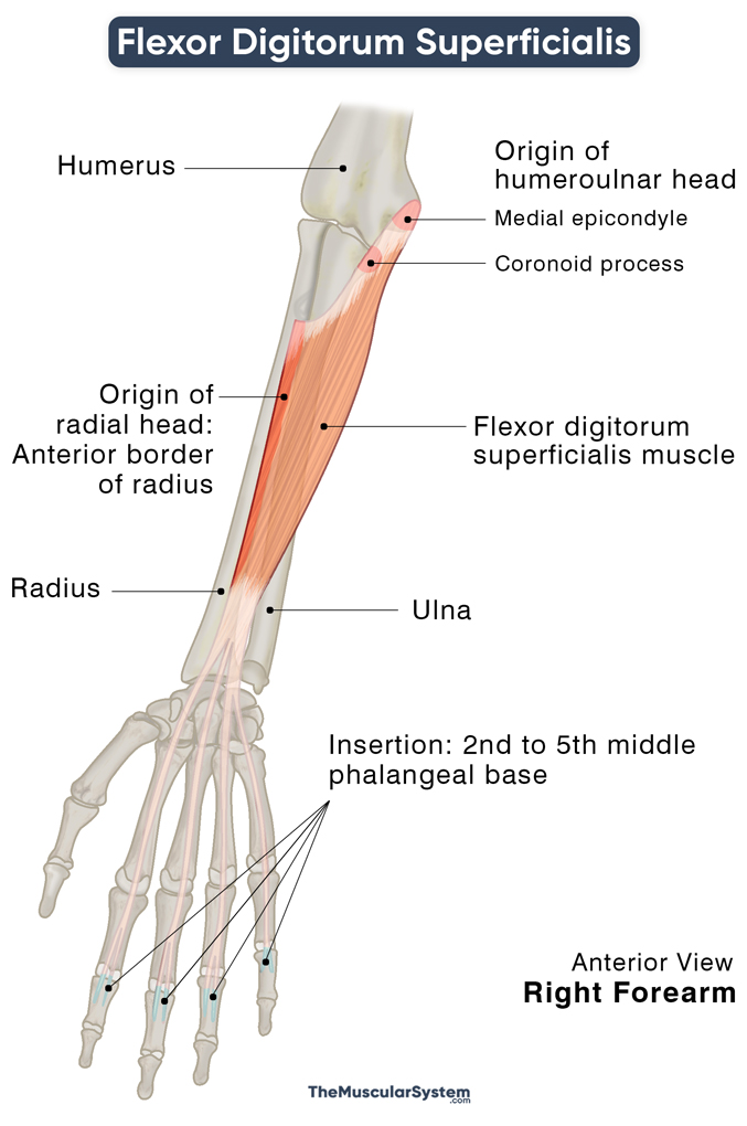
| Origin | Humeroulnar head: Medial epicondyle of the humerus (the common flexor tendon), coronoid process of the ulna, ulnar collateral ligament Radial head: The upper half of the radius’s anterior border |
| Insertion | Bases of the 2nd to 5th middle phalanges |
The first, the humeroulnar head, is named after its two points of origin on the humerus and ulna. Part of it originates from the common flexor tendon, where all the superficial flexors of the forearm originate. The other part rises from the coronoid process, mainly from its medial margin, with a few fibers rising from the ulnar collateral ligament at the elbow joint as well.
The radial head has a linear origin along the anterior border of the radius, from the radial tuberosity extending to the starting point of the pronator tuberosity (point of insertion of the pronator teres).
The two heads join each other through a muscular arch and then continue distally towards the wrist and hand in two distinct muscular layers — superficial and deep. The superficial layer gives off two tendons that reach the middle phalanges of the 3rd and fourth digits, the middle and ring fingers. The muscle fibers in the deep plane also give rise to two tendons directed to the 2nd and 5th digits, or the index and small fingers.
The deep layer also has a small slip to join all 4 deep and superficial tendons so they can together pass into the carpal tunnel, where the flexor retinaculum superficial is to them. These make up 4 of the 9 carpal tunnel tendons.
Through the carpal tunnel, the muscle continues into the palm. Its tendinous slips cross the 2nd to 5th metacarpophalangeal joints to insert into the bases of the 2nd to 5th middle phalanges on the hand’s palmar surface.
Relations With Surrounding Muscles and Structures
The flexor digitorum superficialis lies deep to all the other 4 muscles in the anterior compartment: the flexor carpi radialis, pronator teres, palmaris longus, and flexor carpi ulnaris. It is superficial to the deep anterior forearm muscles flexor pollicis longus and flexor digitorum profundus.
The median nerve and the ulnar artery pass through the muscular arch between the humeroulnar and radial heads.
Functions
| Action | Flexion of 2nd to 5th proximal interphalangeal joints, and of the proximal phalanges at the 2nd to 5th metacarpophalangeal joints |
Primary Function — Flexing the Fingers at Proximal Interphalangeal Joints
Like most muscles in the human body, flexor digitorum superficialis is named after its primary function: flexing the 2nd to 5th digits. When you make a fist, or grab something with your hands, the fingers fold toward the palm with the help of this muscle. This action does not include the thumb, which flexes using other muscles.
To be more specific, the muscle flexes the 2nd to 5th middle phalanges at their respective proximal interphalangeal joints. It is one of the two long finger flexors, the other being the flexor digitorum profundus, which flexes the fingers slowly, and all at once.
The flexor digitorum superficialis flexes the fingers quickly and helps flex them against resistance. Since it is attached to each of the 2nd to 5th middle phalanx individually, it flexes each of the four joints independently, allowing you to bend each finger individually.
Additional Functions
- The muscle also helps flex the 2nd to 5th digits at their metacarpophalangeal joints, which allows you to bend the fingers at their base without curling them up.
- Lastly, flexor digitorum superficialis also helps with wrist flexion.
The primary antagonist of flexor digitorum superficialis is the superficial posterior forearm muscle extensor digitorum.
Innervation
| Nerve | Median nerve (C7, C8 and T1) |
The median nerve (C7, C8, and T1) that innervates the flexor digitorum superficialis is a branch of the brachial plexus. The skin area overlying the muscle receives nerve supply from the roots C6, C7, C8, and T1.
Blood Supply
| Artery | Ulnar artery |
The muscle’s primary blood supply comes from the ulnar artery. In addition to that, its anterior and lateral surfaces receive additional blood supply from the muscular branches of the radial artery. Similarly, the posterior surface receives supplementary blood supply from the muscular branches of the median artery.
References
- Flexor Digitorum Superficialis Muscle: RadioPaedia.org
- Anatomy, Shoulder and Upper Limb, Hand Flexor Digitorum Superficialis Muscle:NCBI.NLM.NIH.gov
- Flexor Digitorum Superficialis Muscle: Muscle: KenHub.com
- Flexor Digitorum Superficialis Muscle: ScienceDirect.com
- Flexor Digitorum Superficialis Muscle: GetBodySmart.com
- Flexor Digitorum Superficialis: IMAIOS.com
- Flexor Digitorum Superficialis: Rad.Washington.edu
Della Barnes, an MS Anatomy graduate, blends medical research with accessible writing, simplifying complex anatomy for a better understanding and appreciation of human anatomy.
- Latest Posts by Della Barnes, MS Anatomy
-
Tensor Tympani
- -
Stapedius
- -
Auricularis Posterior
- All Posts


