

Flexor Pollicis Longus
By
Della Barnes, an MS Anatomy graduate, blends medical research with accessible writing, simplifying complex anatomy for a better understanding and appreciation of human anatomy.
Last updated:
05/05/2023Della Barnes, MS Anatomy
UX/UI Designer at - AdobeDella Barnes, an MS Anatomy graduate, blends medical research with accessible writing, simplifying complex anatomy for a better understanding and appreciation of human anatomy.
What is Flexor Pollicis Longus
Flexor pollicis longus (FPL) is one of the deep anterior forearm muscles that form the upper of the two deep muscular layers. The other two deep muscles in the anterior compartment are the flexor digitorum profundus and the pronator quadratus.
The FPL originates in the forearm, with its insertion point lying in the hand, making it an example of an extrinsic hand muscle. It plays a vital role in the flexion of the thumb.
Anatomy
Location and Attachments
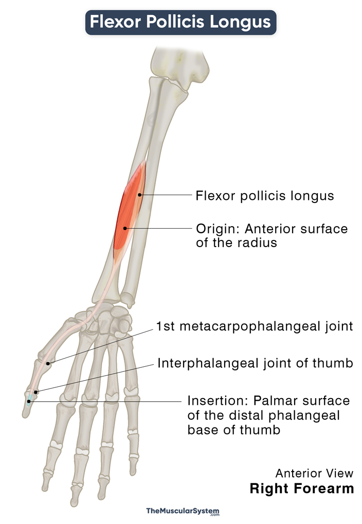
| Origin | The anterior surface of the radius bone and the adjoining interosseous membrane |
| Insertion | The palmar surface of the distal phalangeal base of the thumb |
Origin
Flexor pollicis longus has a broad origin, spanning almost the entire length of the radius’s anterior surface — the palmar side of the forearm. Proximally, it starts from below the radial tuberosity, extending to the area on the distal anterior surface where the pronator quadratus inserts. The point of origin often stretches onto the anterior surface of the adjacent interosseous membrane.
The origin might even continue further to the lateral border of the ulna’s coronoid process.
Sometimes the muscle may have an accessory head of origin arising from the medial side of the coronoid process or the humerus’s medial epicondyle. It happens as a small slip of muscle referred to as the Gantzer muscle.
Insertion
The muscle then travels distally, tapering into a flat, long tendon that passes deep to the flexor retinaculum to enter the carpal tunnel. The tendon passes between the two sesamoid bones lying on the palmar surface in front of the first (thumb) metacarpophalangeal joint. It then inserts into the base of the distal phalanx of the thumb.
Relations With Surrounding Muscles and Structures
At its origin, the flexor pollicis longus lies distal to the deep posterior forearm muscle supinator. It is also radial or lateral to the radial head of flexor digitorum superficialis.
It is one of the three anterior forearm flexors, laterally next to the flexor digitorum profundus and superficial to the pronator quadratus. As already mentioned, it forms the upper layer of the deep anterior forearm muscles along with the flexor digitorum profundus. The groove created by these two muscles lets the anterior interosseous nerve, artery, and vein travel distally toward the hand.
On its way to the carpal tunnel, the FPS tendon travels between the hand muscles opponens pollicis and the adductor pollicis (oblique head) as it courses from beneath the flexor retinaculum.
In some people, the muscle fibers of flexor pollicis longus may blend with those of the flexor digitorum profundus, flexor digitorum superficialis, and pronator teres.
Functions
| Action | Flexion of the thumb |
At the Interphalangeal Joint
It is one of the primary flexors of the thumb and the only muscle that flexes the first interphalangeal joint, helping the thumb flex at this joint. So, the muscle is instrumental in gripping something or picking something up with your fingers and thumb.
At the Metacarpophalangeal Joint
The muscle plays a vital role in flexing the thumb at its metacarpophalangeal joint, which helps stabilize the hand’s grip. You can easily palpate or feel the muscle on the palmar surface of the wrist when you flex your thumb.
The FPL helps with flexing the hand at the wrist joint as well.
The primary antagonists of the flexor pollicis longus are the deep posterior forearm muscles extensor pollicis longus and brevis.
Innervation
| Nerve | Anterior interosseous nerve (C8 and T1) |
The anterior interosseous nerve (C8 and T1) that innervates the muscle is a medial nerve branch with its roots in the spinal nerves.
Blood Supply
| Artery | Anterior interosseous artery |
There are two nerves to supply to two different parts of the muscle. The primary blood supply comes from the ulnar artery branch, the anterior interosseous artery, which supplies the muscle’s medial part. The lateral part receives arterial supply from the radial artery.
A well-developed flexor pollicis longus may receive a third supply from the median artery.
References
- Anatomy, Shoulder and Upper Limb, Hand Flexor Pollicis Longus Muscle: NCBI.NLM.NIH.gov
- Flexor Pollicis Longus: TeachMeAnatomy.info
- Flexor Pollicis Longus Muscle: KenHub.com
- Flexor Pollicis Longus Muscle: RadioPaedia.org
- Flexor Pollicis Longus: IMAIOS.com
- Flexor Pollicis Longus: Rad.Washington.edu
Della Barnes, an MS Anatomy graduate, blends medical research with accessible writing, simplifying complex anatomy for a better understanding and appreciation of human anatomy.
- Latest Posts by Della Barnes, MS Anatomy
-
Tensor Tympani
- -
Stapedius
- -
Auricularis Posterior
- All Posts


