Anterior Forearm Muscles (Forearm Flexors)
Last updated:
19/01/2024Della Barnes, an MS Anatomy graduate, blends medical research with accessible writing, simplifying complex anatomy for a better understanding and appreciation of human anatomy.
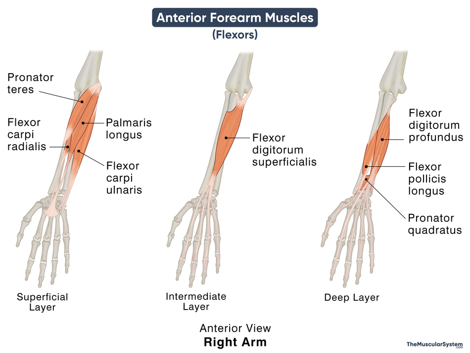
The forearm (antebrachium) is the part of the arm between the elbow and the wrist. All the muscles in the forearm are divided into two compartments, the anterior and posterior. The anterior compartment contains all the muscles at the front of the forearm, that is, the side of the palm. The 8 muscles in the anterior compartment all act to flex the forearm and hand and thus are also known as the forearm flexors.
These muscles lay in different layers within the anterior aspect of the forearm, further classified into superficial, intermediate, and deep groups.
| Muscle Name | Location Layer | Origin | Insertion | Action | Innervation |
|---|---|---|---|---|---|
| 1. Pronator teres | Superficial | Medial epicondyle of the humerus via the common flexor tendon; The coronoid process of ulna | Lateral surface of radius | Pronating the forearm; Flexing the wrist | Median nerve (C6, C7) |
| 2. Flexor carpi radialis | Superficial | Via common flexor tendon | 2nd and 3rd metacarpal base | Flexing and abducting the wrist | Median nerve (C6, C7) |
| 3. Palmaris longus | Superficial | Via common flexor tendon | Into the flexor retinaculum on its distal side | Tightening the grip of hand; Flexing the wrist | Median nerve (C6, C7) |
| 4. Flexor carpi ulnaris | Superficial | Via common flexor tendon; The olecranon of ulna | Pisiform and hamate (carpal bones), and 5th metacarpal base | Flexing and abducting the wrist | Ulnar nerve (C7, C8, T1) |
| 5. Flexor digitorum superficialis | Intermediate | Via common flexor tendon; The coronoid process of ulna; Anterior border of proximal radius | 2nd to 5th middle phalangeal bases | Flexing the index finger to little finger | Median nerve (C6, C7, T1) |
| 6. Flexor digitorum profundus | Deep | Anterior and medial surfaces of proximal ulna; Interosseous membrane | 2nd to 5th distal phalangeal bases | Flexing the index finger to little finger, and the wrist (secondary) | Median nerve (C8 and T1); Ulnar nerve (C8 and T1) |
| 7. Flexor pollicis longus | Deep | Anterior surface of radius; Interosseous membrane | Base of the distal phalanx of the thumb | Flexing the thumb | Anterior interosseous nerve (C8, T1) |
| 8. Pronator quadratus | Deep | Anterior surface of distal ulna | Anterior surface of distal radius | Pornating the forearm | Anterior interosseous nerve (C8, T1) |
All the muscles in the flexor group receive their blood supply from various branches of the ulnar and radial arteries.
FAQs
Ans. Interestingly, the strongest flexor of the forearm at the elbow is the brachialis, which is an upper arm muscle. The flexors in the anterior compartment primarily work at the wrist and hand, with the strongest flexor of the wrist joint being the flexor carpi ulnaris.
Ans. As evident from the table above, most of the muscles in the forearm flexor group are innervated by the median nerve, with help from the ulnar nerve.
References
- Muscles in the Anterior Compartment of the Forearm: TeachMeAnatomy.info
- Superficial Anterior Forearm Muscles: KenHub.com
- Deep Anterior Forearm Muscles: KenHub.com
- Muscles of the Anterior Forearm – Superficial View: LearnMuscles.com
- Anterior Compartment of the Forearm: RadioPaedia.org
Della Barnes, an MS Anatomy graduate, blends medical research with accessible writing, simplifying complex anatomy for a better understanding and appreciation of human anatomy.
- Latest Posts by Della Barnes, MS Anatomy
-
Temporoparietalis
- -
Corrugator Supercilii
- -
Depressor Supercilii
- All Posts


