Brachialis Muscle
Last updated:
18/04/2024Della Barnes, an MS Anatomy graduate, blends medical research with accessible writing, simplifying complex anatomy for a better understanding and appreciation of human anatomy.
What is the Brachialis
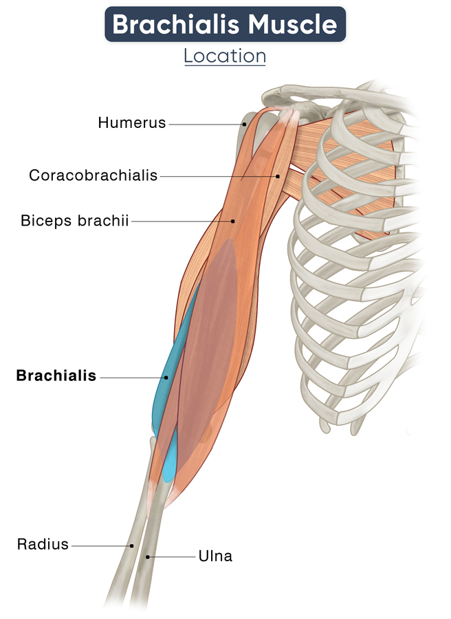
The brachialis is one of the essential muscles in the upper arm, located in the arm’s anterior compartment. It is the muscle primarily responsible for allowing the elbow to flex.
Location
| Origin | Anterior surface of the humerus, mainly on the distal half |
| Insertion | The ulnar tuberosity and the depression on the front surface of the ulna’s coronoid process |
The muscle is located low in the anterior compartment. It is attached to the intermuscular septa of the arm on both sides, with a stronger attachment to the medial intermuscular septum.
The brachialis muscle fibers converge to form a strong tendon, called the brachialis tendon, which then inserts into the ulnar tuberosity and coronoid process. This tendon partially forms the floor of the cubital fossa.
The biceps brachii muscle is situated just above the brachialis, along with the musculocutaneous and median nerves as well as the brachial vessels. Behind it are the humerus bone’s proximal part and the elbow joint capsule.
On the medial side, the medial intermuscular septum and the pronator teres separate the muscle from the ulnar nerve and the triceps brachii.
Anatomical Variation: Usually, there is one brachialis muscle in each arm. But some cases have been recorded where additional accessory muscles are present. These insert into the common flexor tendon at the elbow joint.
Functions
| Action | Flexing the forearm at the elbow joint |
It works with the brachioradialis and biceps brachii muscles to flex and move the upper arm and fold the arm at the elbow. Being the primary flexor of the elbow, the muscle allows it to bend at all physiologic angles. However, it is not as strong when the elbow is in supination because then, the biceps is stronger, making it the primary flexor.
The brachialis has no attachment to the radius, so it is not involved in pronation and supination.
Because of its superficial location and greater volume, the superficial head of the brachialis is mechanically more suitable to stabilize the arm in flexion. It is at its strongest at a 90-degree flexion of the arm.
On the other hand, the smaller or deep head helps initiate flexing of the elbow when the arm is outstretched.
Innervation
| Nerve | Musculocutaneous nerve (C5, C6); Radial nerve |
The musculocutaneous nerve, with its roots in the cervical nerves C5 and C6, innervates the muscle medially. It runs superficially on the muscle’s surface, between the brachialis and biceps brachii.
In most people, this muscle has dual innervation with the radial nerve (C5-T1) also innervating it laterally.
Blood Supply
| Artery | Muscular branches of brachial artery and radial recurrent artery |
Occasionally, the inferior and superior ulnar collateral arteries can also supply this muscle.
References
- Brachlialis Muscle – Location & Function – Study.com
- Brachialis – Osmosis.org
- Anatomy, Shoulder and Upper Limb, Brachialis Muscle – Ncbi.nlm.nih.gov
- Brachialis muscle – Kenhub.com
- Brachialis Muscle – Getbodysmart.com
Della Barnes, an MS Anatomy graduate, blends medical research with accessible writing, simplifying complex anatomy for a better understanding and appreciation of human anatomy.
- Latest Posts by Della Barnes, MS Anatomy
-
Tensor Tympani
- -
Stapedius
- -
Auricularis Posterior
- All Posts


