

Brachioradialis
By
Della Barnes, an MS Anatomy graduate, blends medical research with accessible writing, simplifying complex anatomy for a better understanding and appreciation of human anatomy.
Last updated:
05/05/2023Della Barnes, MS Anatomy
UX/UI Designer at - AdobeDella Barnes, an MS Anatomy graduate, blends medical research with accessible writing, simplifying complex anatomy for a better understanding and appreciation of human anatomy.
What is Brachioradialis
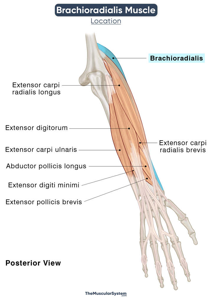
The brachioradialis is a fusiform superficial muscle in the posterior compartment of the forearm. It forms the ‘mobile wad’ or radial group of forearm muscles along with extensor carpi radialis brevis and extensor carpi radialis longus. Named for its location and action, it runs from the humerus’s base to the radius’s base.
Despite being one of the 6 superficial posterior muscles of the forearm, where all the muscles act as forearm extensors, the brachioradialis works on flexing the forearm, mainly due to the orientation of its fibers. The other 5 muscles in this compartment are the extensor digitorum, extensor digiti minimi, extensor carpi ulnaris, and extensor carpi radialis longus and brevis.
Anatomy
Location and Attachments
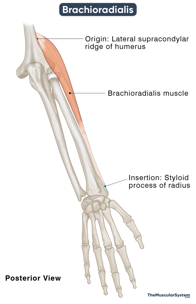
| Origin | Lateral supracondylar ridge of the humerus |
| Insertion | Styloid process on the distal side of the radius |
Origin
The muscle originates from the proximal side of the humerus, on the upper two-thirds of the lateral supracondylar ridge. Some fibers also originate from the anterior side of the arm’s lateral intermuscular septum — the boundary between the anterior and posterior compartments of the forearm.
The muscle courses distally, passing over the lateral surface of the elbow joint. Here, it enters the region of the cubital fossa, with its medial surface forming the lateral wall of the fossa. The cubital fossa is the triangular depression on the inner side of the elbow where the median and radial nerves, brachial artery, and biceps tendon lie.
Insertion
The brachioradialis fibers continue further down, through the anterior side of the forearm on the radial side. The muscle belly fibers converge to form a thick tendon around the middle third of the forearm, which runs down towards the wrist joint to insert into the radius’s styloid process.
The brachioradialis is a unique muscle with its origin on the distal end of one bone and its insertion in the distal end of another bone.
Relations With Surrounding Muscles and Structures
It contributes to the muscle mass that forms the anterior-lateral side of the elbow joint. Since it is a superficial muscle, it lies directly beneath the skin layer, overlying all the other muscles in the area. The muscle can easily be palpated on the elbow’s outer (radial) side, especially in a flexed position, with the forearm partially pronated.
The biceps brachii runs deep to the brachioradialis as it inserts into the radial tuberosity. Proximally, it has the distal end of the brachialis muscle on its medial side. The space between the brachioradialis and brachialis allows passage to the radial nerve and the arterial anastomosis of the elbow (between deep brachial and radial recurrent arteries).
The flexor carpi ulnaris is located medial to the brachioradialis around the middle of the forearm.
The lateral cutaneous antebrachial nerve and the cephalic vein are superficial to the muscle, running over its superior surface.
On the distal side, the radial artery lies on the ulnar side of the muscle’s tendon, marking the spot where the radial pulse is taken. The radial nerve’s superficial branch rises here, deep to the brachioradialis. Like on its proximal side, the muscle lets the radial nerve pass through the space between itself and the extensor carpi radialis brevis.
Just before the tendon inserts into the styloid process, it is crossed by the inserting tendons of the deep forearm muscles, abductor pollicis longus, and extensor pollicis brevis.
Function
| Action | Flexion of the forearm and the elbow joint (primary), stabilizing the elbow (secondary), pronation of the forearm (secondary) |
1. Flexing the Forearm
The primary action of brachioradialis is working with the brachialis and biceps brachii as a synergistic to bend the arm at the elbow joint.
All these three muscles are active during flexion, whether the arm is pronated, neutral, or supinated. But their attachments and the orientation of their fibers decide which one will be the primary flexor when the forearm is in a specific position.
As mentioned above, brachioradialis is unique in its proximal attachment at the distal end of the humerus before the elbow joint and its distal attachment at the distal end of the radius near the wrist joint. These two attachments enable the muscle to work like a lever, where the elbow joint acts like the fulcrum. The muscle works as the primary flexor when the forearm is semi-pronated, having the palm perpendicular to the ground.
This movement is key to most arm functions, including putting food into your mouth, holding the phone to your ear, and rowing.
2. Pronating/Supinating the Forearm
The brachioradialis always tries to keep the forearm in a semi-pronated position. When the forearm is pronated, the muscle supinates it to a 90° position so it can be semi-pronated again. Similarly, it pronates the forearm from a supinated position to reach the semi-pronated stance.
3. Stabilizing the Elbow Joint
Along with the biceps brachii and brachialis, the brachioradialis works to keep the elbow joint and forearm stable during flexion, pronation, and supination.
The antagonists of the brachioradialis are the triceps brachii and anconeus.
Innervation
| Nerve | Radial nerve (C5, and C6) |
The radial nerve, with nerve roots C5 and C6, innervates this muscle. It is a branch of the brachial plexus via its posterior cord.
Blood Supply
| Artery | Radial recurrent artery |
The primary blood supply comes from the radial recurrent artery, a branch of the radial artery. Another source of blood supply is the radial collateral artery, a deep brachial artery branch.
References
- Brachioradialis Muscle: Function & Nerve: Study.com
- Brachioradialis Muscle: KenHub.com
- Brachioradialis: Osmosis.org
- Brachioradialis Muscle:IMAIOS.com
- Brachioradialis Muscle: RadioPaedia.org
- Anatomy, Shoulder and Upper Limb, Forearm Brachioradialis Muscle: NCBI.NLM.NIH.gov
- Brachioradialis Muscle – Attachments, Action & Innervation: GetBodySmart
Della Barnes, an MS Anatomy graduate, blends medical research with accessible writing, simplifying complex anatomy for a better understanding and appreciation of human anatomy.
- Latest Posts by Della Barnes, MS Anatomy
-
Tensor Tympani
- -
Stapedius
- -
Auricularis Posterior
- All Posts


