Extensor Carpi Ulnaris
Last updated:
04/06/2024Della Barnes, an MS Anatomy graduate, blends medical research with accessible writing, simplifying complex anatomy for a better understanding and appreciation of human anatomy.
What is Extensor Carpi Ulnaris
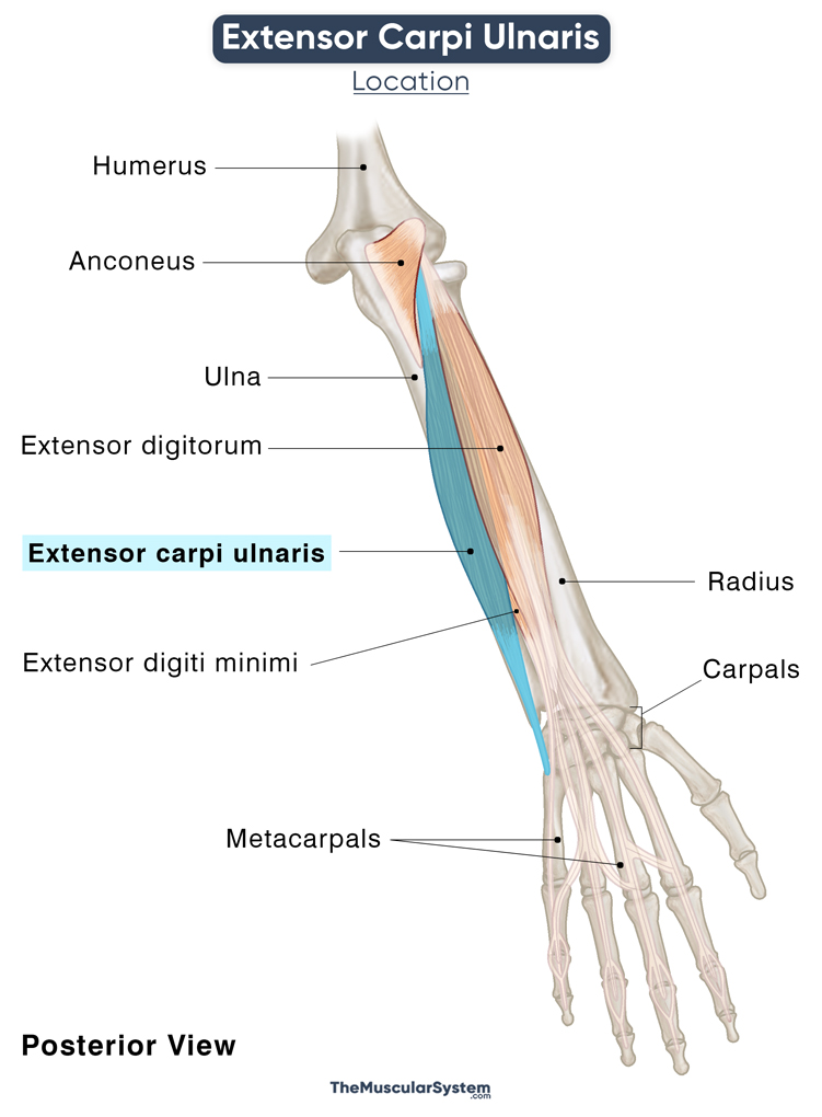
The Extensor carpi ulnaris (ECU) is a long fusiform muscle in the forearm, mainly responsible for wrist movements. It is one of the superficial muscles in the forearm’s posterior compartment, along with the extensor digitorum, extensor digiti minimi, brachioradialis, and the extensor carpi radialis longus and brevis (and the anconeus).
Anatomy
Location and Attachments
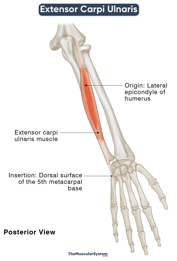
| Origin | Lateral epicondyle of the humerus (the common extensor tendon) and posterior border of the ulna |
| Insertion | The dorsal surface of the 5th metacarpal base |
Origin
The muscle originates via two heads, the humeral and ulnar heads. The humeral head arises from the humerus’s lateral epicondyle via the common extensor tendon, an origin that it shares with the extensor digitorum, extensor digiti minimi, and extensor carpi radialis brevis. A few fibers also originate from the fascia adjacent to the extensor tendon.
The ulnar head originates from the posterior ulnar border via the common aponeurosis and the forearm’s deep fascia. The common extensor tendon that arises from the common aponeurosis also serves as the origin point of flexor digitorum profundus and flexor carpi ulnaris.
Insertion
The muscle belly then courses medially and downward towards the ulnar side of the forearm. It narrows into a tendon just above the wrist joint, which then passes through the groove between the posterior surface of the ulnar head, and its styloid process.
The ligament courses individually through a synovial sheath, forming the 6th (the most medial) compartment of the extensor retinaculum. It then enters the hand’s dorsal surface, travels superficially to the carpal bone triquetrum, and inserts into the base of the 5th metacarpal (of the little finger).
Relations With Surrounding Muscles and Structures
Since it is the most medially located of all the posterior forearm muscles, the retinaculum compartment containing the extensor carpi ulnaris only has the elbow muscle, anconeus, lying medial to it. The extensor digitorum and extensor digiti minimi are lateral to it.
There is a passage between the extensor and flexor carpi ulnaris muscles, at the distal end, just before the wrist joint. It is where the dorsal cutaneous branch of the ulnar artery originates.
Function
| Action | Extension and adduction of the hand at the wrist joint |
Extending the Wrist
The extensor carpi ulnaris works with two other superficial posterior forearm muscles, extensor carpi radialis longus, and brevis, to extend the wrist joint. It means bending the wrist to have your palm face out — like you would do to stop a bus.
Additionally, it helps stabilize the fist and clench it tightly when you grab something. When your wrist is extended, it blocks the action of the forearm flexors on the wrist. As a result, these flexors only act on the digits, helping strengthen the grip. An example of such a grip with an extended wrist would be when you hit a backhand at tennis.
Adducting the Wrist
The muscle also works along the flexor carpi ulnaris for adduction of the hand at the wrist joint, which means bending the wrist toward the ulnar side (on the side of the little finger). Since one is a flexor, and the other is an extensor, the synergistic action of the two ensures there is no added flexion or extension during this ulnar adduction. This kind of movement is essential for things like swinging a baseball bat or hammering a nail.
The extensor carpi ulnaris also helps keep the distal radioulnar joint stable.
The primary antagonist of the extensor carpi ulnaris is the superficial anterior forearm muscle flexor carpi radialis.
Innervation
| Nerve | Posterior interosseous nerve (C7, and C8) |
The posterior interosseous nerve, having its roots in the spinal nerves C7 and C8, and arising from the radial nerve’s deep division, innervates the muscle.
Blood Supply
| Artery | Ulnar artery, and posterior interosseous artery |
The Extensor carpi ulnaris gets its primary blood supply from the ulnar artery. Additional vasculature comes from the posterior interosseous artery, which branches off the radial artery and supplies both the superficial and deep muscle groups of the forearm’s posterior compartment.
References
- Extensor Carpi Ulnaris Muscle: KenHub.com
- Extensor Carpi Ulnaris: Insertion, Innervation, & Action: Study.com
- Extensor Carpi Ulnaris Muscle: RadioPaedia.org
- Extensor Carpi Ulnaris: IMAIOS.com
- Extensor Carpi Ulnaris: Rad.Washington.edu
- Extensor Carpi Ulnaris – Attachments, Action & Innervation: GetBodysmart.com
Della Barnes, an MS Anatomy graduate, blends medical research with accessible writing, simplifying complex anatomy for a better understanding and appreciation of human anatomy.
- Latest Posts by Della Barnes, MS Anatomy
-
Infrahyoid Muscles
- -
Omohyoid
- -
Sternohyoid
- All Posts


