Flexor Carpi Radialis
Last updated:
04/06/2024Della Barnes, an MS Anatomy graduate, blends medical research with accessible writing, simplifying complex anatomy for a better understanding and appreciation of human anatomy.
What is Flexor Carpi Radialis
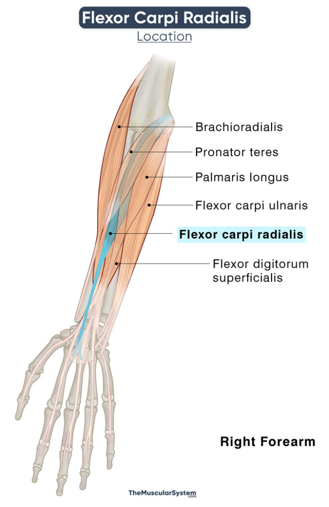
The flexor carpi radialis, or FCR, is a thin long muscle in the anterior compartment of the forearm. It is one of the five superficial muscles in the forearm, with the others being pronator teres, palmaris longus, flexor carpi ulnaris, and flexor digitorum superficialis. The muscle runs from the humerus to the second metacarpal bone, having a vital role in the movement of the wrist and hand.
Anatomy
Location and Attachments
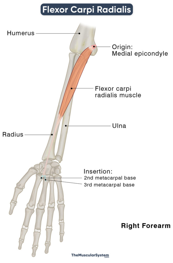
| Origin | Medial epicondyle of the humerus (the common flexor tendon) |
| Insertion | The 2nd and 3rd metacarpal bases |
Flexor carpi radialis originates from the common flexor tendon at the medial epicondyle of the elbow. The other four superficial muscles of the anterior compartment also originate from the same tendon, hence the name ‘common’ flexor tendon. The muscle belly courses obliquely, crossing to the radial side (thumb-side) of the arm from the ulnar side (side of the small finger) where it originated.
It then turns into a tendon in the lower third region of the forearm, passing into the surface of the palm from underneath the flexor retinaculum, which is the connective tissue band forming the anterior wall of the carpal tunnel. This attaching tendon constitutes almost half the total length of the muscle. A synovial sheath forms a tunnel in the flexor retinacular space to allow passage exclusively to this tendon, letting it pass the scaphoid bone and the groove on the trapezium bone on the palmar surface.
Upon reaching the metacarpals, the tendon divides into two to insert into the bases of the 2nd and 3rd metacarpals.
Relations With Surrounding Muscles and Structures
Since it is a superficial muscle, it is deep only to the fat tissue and skin layer, with the flexor digitorum superficialis muscle lying deep to it. The flexor carpi radialis is located between the pronator teres on its lateral side and the palmaris longus on its medial side. It lies medial to the posterior compartment muscle brachioradialis at its posterior side.
At its insertion point, the tendon fibers are deep to the oblique head of the hand muscle adductor pollicis.
Flexor carpi radialis is also superficial to a couple of important neurovascular structures. The median nerve runs from beneath the proximal side of the muscle right before it passes underneath the flexor digitorum superficialis.
On the inner side of the wrist, the radial artery courses from between the flexor carpi radialis and brachioradialis muscle tendons. The artery can be palpated at this point, along with the tendon of flexor carpi radialis. It is where the doctors take your (radial) pulse at the wrist.
Functions
| Action | Flexing and abducting the hand at the wrist |
Flexing the wrist means bending it forward, or towards the inside of the hand. Whereas abducting the wrist means bending it sideways, on the side of the thumb. The movements are crucial for amost all daily activities like eating, brushing the teeth, driving, and typing.
As mentioned above, the muscle runs obliquely, crossing the width of the forearm from the medial epicondyle on the ulnar side of the forearm to the 2nd and 3rd metacarpals on the radial side. This connection between its origin and insertion means it can pull the hand both proximally and laterally, producing both flexing and abducting movements.
It works with the palmaris longus and flexor carpi ulnaris to allow the wrist to flex without abducting. Then again, it is synergistic to the extensor carpi radialis brevis and longus muscles. And the counteracting forces of these two actions result in a balanced abduction of the wrist, which means abduction without flexion.
Among its minor functions are contribtuing to the pronation of the hand, and preventing excessive extension and possible displacement of the hand when the fingers are extended.
The primary antagonist of the flexor carpi ulnaris is the posterior compartment muscle extensor carpi ulnaris.
Innervation
| Nerve | Median nerve (C6, and C7) |
The median nerve (C6, and C7), its major source of innervation, is a branch of the brachial plexus — its medial and lateral cords to be more specific.
Blood Supply
| Artery | Either the anterior or posterior recurrent ulnar artery |
The primary blood supply comes from a branch of either the anterior or the posterior recurrent ulnar arteries in the forearm near the elbow joint. The muscle receives additional blood supply from 6-8 radial artery branches.
References
- Flexor Carpi Radialis: IMAIOS.com
- Anatomy, Shoulder and Upper Limb, Forearm Flexor Carpi Ulnaris Muscle: StatPearls.com
- Flexor Carpi Radialis Muscle: KenHub.com
- Flexor Carpi Radialis: Definition, Function & Nerve: Study.com
- Flexor Carpi Radialis: Rad.Washington.edu
- Flexor Carpi Radialis Muscle: ScienceDirect.com
- Flexor Carpi Radialis: GetBodySmart.com
Della Barnes, an MS Anatomy graduate, blends medical research with accessible writing, simplifying complex anatomy for a better understanding and appreciation of human anatomy.
- Latest Posts by Della Barnes, MS Anatomy
-
Laryngeal Muscles
- -
Thyroarytenoid
- -
Lateral Cricoarytenoid
- All Posts


