

Dorsal Interossei of the Foot
By
Della Barnes, an MS Anatomy graduate, blends medical research with accessible writing, simplifying complex anatomy for a better understanding and appreciation of human anatomy.
Last updated:
28/10/2025Della Barnes, MS Anatomy
UX/UI Designer at - AdobeDella Barnes, an MS Anatomy graduate, blends medical research with accessible writing, simplifying complex anatomy for a better understanding and appreciation of human anatomy.
What Are the Dorsal Interossei of the Foot
The dorsal interossei are a group of four bipennate, intrinsic muscles located between the metatarsal bones of the foot. It is one of the two muscles in the fourth and deepest layer of plantar muscles, with the other being the plantar interossei.
From medial to lateral, the four muscles are numbered as the first, second, third, and fourth dorsal interosseous. Their functions include flexing, abducting, and extending the toes.
Anatomy
Location and Attachments
| Origin | The sides of the proximal half of the 1st-5th metatarsal bones |
| Insertion | The bases of the 2nd-4th proximal phalanges of the lateral toes |
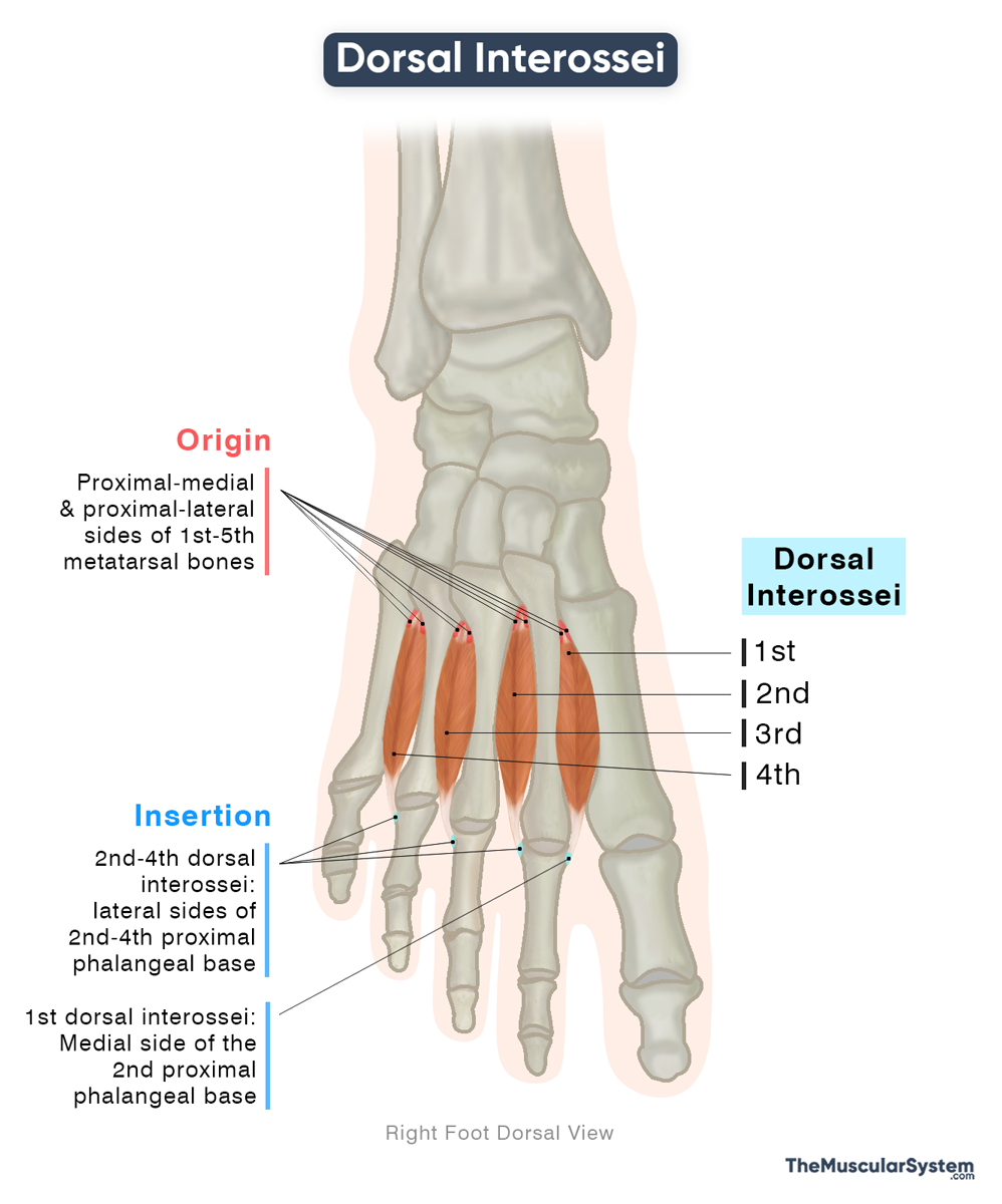
Origin
The muscles originate from both the medial and lateral sides of the proximal half of the first to 5th metatarsal bones. Each dorsal interosseous has two heads, originating from the medial and lateral sides of the two metatarsals between which it lies. So, the first dorsal interosseous arises from the medial side of the first metatarsal and the lateral side of the second metatarsal, and so on. This gives the muscles their bipennate or feather-like shape.
Insertion
The two heads from the opposite sides converge and form a narrow tendon for each dorsal interosseous. So, there are a total of four tendons, coursing forward toward the phalanges. The first, most medial tendon inserts into the medial side of the second proximal phalangeal base. The other three tendons from the second to fourth dorsal interossei insert into the lateral side of the second to fourth proximal phalangeal bases. Some part of the tendon also inserts into the adjacent extensor hoods.
So, the second proximal phalanx has two dorsal interossei inserting into it, one on its medial side and the other on its lateral side.
Relations With Surrounding Muscles and Structures
Belonging to the deepest layer of plantar muscles, the dorsal interossei lie immediately deep to the third-layer muscles, the flexor hallucis brevis, adductor hallucis, and flexor digiti minimi brevis. Each of the four dorsal interossei also lies superficial to the corresponding lumbrical muscles of the second layer.
Originating from the adjacent sides of the metatarsals, the medial and lateral heads of each dorsal interosseous muscle incline slightly forward and toward one another. This arrangement creates small angular gaps in the upper parts of the intermetatarsal spaces, through which the following blood vessels pass from the dorsum of the foot into the sole.
- The deep plantar branch of the dorsalis pedis artery passes between the two heads of the first dorsal interosseous to contribute to the plantar arterial arch.
- The posterior perforating arteries pass between the two heads of each of the second and fourth dorsal interossei.
Function
| Action | — Flexion and abduction of the 2nd-4th digits at the metatarsophalangeal joints — Extension of the 2nd-4th digits at the interphalangeal joints |
Despite their small size, these muscles play a vital role in controlling the second, third, and fourth toes and maintaining foot stability.
Flexion of the Toes
Working with the plantar interossei, these muscles flex the second to fourth toes at the metatarsophalangeal joints. This flexion helps the flexor digitorum longus and brevis to function more effectively during movements such as running or jumping.
Assistance in Abduction
The muscles also abduct the same toes at the same joints, which means each interosseous pulls its respective toe slightly away from the foot’s midline. Though this is a minor action, it helps with fine motor control and balance during gait.
Extension of the Toes
Another important action of these muscles is assisting the extensor digitorum longus and brevis in extending the same toes at their interphalangeal joints, helping coordinate smooth toe movement.
Stability and Alignment
The second toe forms the anatomical midline of the foot. The first and second dorsal interossei lie on either side of it, pulling in opposite directions to maintain alignment. Collectively, the four muscles support the forefoot and help preserve the medial and lateral arches, enhancing stability during walking, running, and jumping.
Antagonists
The plantar interossei are antagonistic to the dorsal interossei because they adduct the same toes that the dorsal interossei abduct. However, both muscle groups act synergistically during toe flexion.
Innervation
| Nerve | Lateral plantar nerve (S2-S3) |
All four dorsal interosseous muscles are innervated by the lateral plantar nerve, a terminal branch of the tibial nerve that carries fibers from the second and third sacral nerve roots (S2-S3).
Blood Supply
| Artery | Dorsalis pedis, dorsal metatarsal, plantar metatarsal, and lateral plantar arteries |
The muscle receives blood supply from both the anterior and posterior tibial arteries. The anterior tibial artery contributes via the dorsalis pedis and dorsal metatarsal arteries, while the posterior tibial artery supplies the muscle through its lateral plantar and plantar metatarsal branches.
References
- Dorsal Interossei (Foot): Radiopaedia.org
- Dorsal Interossei Muscles of the Foot: Kenhub.com
- Dorsal Interossei (Foot): TeachMeAnatomy.info
- Dorsal Interossei Muscles of Foot (Left): Elsevier.com
- Dorsal Interossei of Feet: WheelessOnline.com
Della Barnes, an MS Anatomy graduate, blends medical research with accessible writing, simplifying complex anatomy for a better understanding and appreciation of human anatomy.
- Latest Posts by Della Barnes, MS Anatomy
-
Tensor Tympani
- -
Stapedius
- -
Auricularis Posterior
- All Posts


