

Dorsal Interossei of Hand
By
Della Barnes, an MS Anatomy graduate, blends medical research with accessible writing, simplifying complex anatomy for a better understanding and appreciation of human anatomy.
Last updated:
30/03/2024Della Barnes, MS Anatomy
UX/UI Designer at - AdobeDella Barnes, an MS Anatomy graduate, blends medical research with accessible writing, simplifying complex anatomy for a better understanding and appreciation of human anatomy.
What are the Dorsal Interossei
The dorsal interossei (DI) are a group of 4 intrinsic hand muscles located between the 2nd to 5th metacarpals. These small muscles are involved in the movements of the fingers.
The muscles are referred to as the 1st to 4th dorsal interosseous from the radial to the ulnar side. You could easily feel them in the dorsum of the hand, between the 2nd to 5th metacarpals, or between the 4 inserting tendons of the extensor digitorum. The flat, triangular 1st dorsal interosseous muscle (also called the abductor indicis) is the largest of the 4 and is the easiest to palpate in the web between the index finger and the thumb.
Anatomy
Dorsal interossei are bipennate in structure, which means the muscle fibers extend in opposite directions from a central tendon, giving the muscle a feather-like appearance. They are the most superficial muscles on the dorsum of the hand and the most dorsally located intrinsic hand muscles.
Location and Attachments
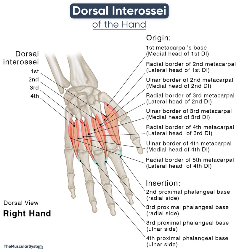
Each of these muscles arise via two heads.
| 1st dorsal interosseous | 2nd dorsal interosseous | 3rd dorsal interosseous | 4th dorsal interosseous | |
|---|---|---|---|---|
| Origin | Medial head: The palmar surface and ulnar side of the 1st or thumb metacarpal’s base Lateral head: The radial border of the 2nd or the index finger’s metacarpal | Medial head: The ulnar border of the 2nd metacarpal Lateral head: The radial border of the 3rd or middle finger’s metacarpal | Medial head: The ulnar border of the 3rd metacarpal Lateral head: The radial border of the 4th or ring finger’s metacarpal | Medial head: The ulnar border of the 4th metacarpal Lateral head: The radial border of the 5th or little finger’s metacarpal |
| Insertion | The radial side of the index finger’s proximal phalangeal base and the adjacent extensor expansion | The radial side of the middle finger’s proximal phalangeal base and the adjacent extensor expansion | The ulnar side of the middle finger’s proximal phalangeal base and the adjacent extensor expansion | The ulnar side of the ring finger’s proximal phalangeal base and the adjacent extensor expansion |
Some structural variations may exist where some of the dorsal interossei may be unipennate or have three heads instead of two. Their point of insertion may sometimes vary as well.
Relations With Surrounding Muscles and Structures
As mentioned, these muscles occupy the interosseous space in the hand’s dorsal compartment. They have the palmar interossei lying opposite them on the palmar side. The inserting tendon of each dorsal interosseous muscle passes from behind the deep transverse metacarpal ligaments to insert into the proximal phalanx of their respective digit.
The two originating heads of each interosseous create a triangular space with the adjacent carpometacarpal joint. Such space created by the 1st interosseous allows passage to the radial artery as it enters the palm from the dorsal side of the hand. Similar spaces created by the rest of the interossei muscles let the perforating branches of the deep palmar arch (radial artery) pass.
Function
| Action | Finger abduction and flexion at the metacarpophalangeal joint; finger extension at the interphalangeal joints |
1. Finger Abduction
The primary function of these muscles is the abduction of the fingers, which means letting them move away from each other when you spread your fingers apart. The 1st and 2nd dorsal interossei cause radial abduction of the index and middle fingers, pulling them laterally toward the thumb. Similarly, the 3rd and 4th dorsal interossei cause ulnar abduction of the ring and little fingers to pull them medially away from the thumb.
Note that the middle finger can abduct laterally as well as medially, depending on whether the 2nd or the 3rd interosseous is contracting. It happens because both the 2nd and 3rd interossei attach to the proximal phalanx of the middle finger.
The abductor pollicis brevis and the abductor digiti minimi muscles assist the dorsal interossei in this function as they abduct the thumb and little finger, respectively. Therefore, the actions of the palmar interossei, which brings the fingers close together, are antagonistic to that of the dorsal interossei.
Mnemonic
PAD DAB (DAB with a Pad)
- Palmar interossei – ADduct
- Dorsal interossei – ABduct
2. Finger Flexion at the Metacarpophalangeal Joints
Along with the abduction, these muscles also help flex the index to little fingers at the metacarpophalangeal joints. Though it is only a minor action, it helps the primary flexors of the fingers, the flexor digitorum superficialis, flexor digitorum profundus, and lumbricals.
3. Finger Extension at the Interphalangeal Joints
In addition to their actions at the metacarpophalangeal joints, these muscles also act on the proximal and distal interphalangeal joints to extend the fingers.
The actions of the dorsal interossei help stabilize the joints in the fingers.
Innervation
| Nerve | Deep branch of ulnar nerve (C8 and T1) |
All the dorsal interossei of the hand are innervated by the deep branch of the ulnar nerve arising from the nerve roots C8 and T1. The part of the skin overlying the muscle receives innervation from the same nerve with nerve roots C6 to C8.
Blood Supply
| Artery | Dorsal metacarpal arteries and palmar metacarpal arteries |
The 1st dorsal interosseous receives primary blood supply from the 1st dorsal metacarpal artery, a branch of the radial artery. The other dorsal interossei are supplied by the 2nd, 3rd, and 4th dorsal metacarpal arteries, branching from the dorsal carpal network.
Additional blood supply comes from the radial, princeps pollicis, and radialis indicis arteries. The perforating branches of the deep palmar arch that pass through the triangular spaces at the bases of these muscles also vascularise them.
References
- Dorsal Interossei Muscles of Hand: IMAIOS.com
- Dorsal Interossei Muscles (Hand): RadioPaedia.org
- Dorsal Interossei (Hand): HealthLine.com
- Anatomy, Shoulder and Upper Limb, Hand Dorsal Interossei Muscle: NCBI.NLM.NIH.gov
- Dorsal Interossei Muscles of the Hand: KenHub.com
- Dorsal Interossei: Meddean.LUC.edu
- Interosseous Muscles, Dorsal: Rad.Washington.edu
Della Barnes, an MS Anatomy graduate, blends medical research with accessible writing, simplifying complex anatomy for a better understanding and appreciation of human anatomy.
- Latest Posts by Della Barnes, MS Anatomy
-
Tensor Tympani
- -
Stapedius
- -
Auricularis Posterior
- All Posts


