

Palmar Interossei
By
Della Barnes, an MS Anatomy graduate, blends medical research with accessible writing, simplifying complex anatomy for a better understanding and appreciation of human anatomy.
Last updated:
14/05/2025Della Barnes, MS Anatomy
UX/UI Designer at - AdobeDella Barnes, an MS Anatomy graduate, blends medical research with accessible writing, simplifying complex anatomy for a better understanding and appreciation of human anatomy.
What are the Palmar Interossei
The palmar interossei or volar interossei are 3 (sometimes 4) small intrinsic hand muscles between the metacarpals on the palmar side of the hand. The palmar interossei are much smaller than the dorsal interossei.
These muscles are the fingers’ primary adductors and help with flexion and extension.
Anatomy
All the muscles in this group are unipennate, originating via a single head, with the muscle fibers coursing obliquely from the point of origin. Though there are usually 3 palmar interossei in a human hand, sometimes there may be 4, with an additional muscle at the base of the thumb.
Location and Attachments
Based on their position, and associated digits, the 4 muscles are named the 1st, 2nd, 3rd, and 4th palmar interossei. Studies have shown the 1st palmar interosseous, also known as the pollical palmar interosseous, to be more commonly present than absent. But, most anatomy texts consider it rudimentary, even when present.
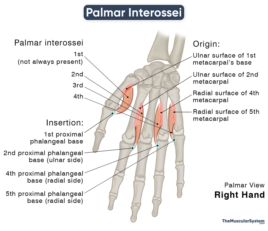
| 1st palmar interosseous | 2nd palmar interosseous | 3rd palmar interosseous | 4th palmar interosseous | |
|---|---|---|---|---|
| Origin | The ulnar surface of the 1st metacarpal’s base | The entire length of the 2nd metacarpal’s ulnar surface | The radial side of the 4th metacarpal | The radial side of the 5th metacarpal |
| Insertion | The 1st proximal phalangeal base and the adjacent extensor expansion | The ulnar side of the 2nd proximal phalangeal base and the adjacent extensor expansion | The radial side of the 4th proximal phalangeal base and the adjacent extensor expansion | The radial side of the 5th proximal phalangeal base and the adjacent extensor expansion |
All the palmar interossei insert into their associated proximal phalanx, on the same side from which they originate. Only the 1st palmar interossei insert into a small sesamoid bone on the ulnar side of the thumb, in addition to the 1st proximal phalangeal base.
The 1st palmar interosseous is often considered a part of the adductor pollicis or flexor pollicis brevis. Studies have suggested that this muscle was initially derived from the adductor pollicis’ oblique head, evolving to become the small individual muscle it is today.
Relations With Surrounding Muscles and Structures
The palmar interossei lie on the palmar side of the hand, opposite to the dorsal interossei. Together, all the interossei muscles fill up the spaces between the 5 metacarpal bones of the hand.
The 1st palmar interosseous, when present, has the 1st dorsal interosseous lying on its dorsal side, with the oblique head of adductor pollicis lying in front of it.
The 2nd palmar interosseous also lies adjacent to the oblique head of adductor pollicis. Similarly, the 3rd and 4th palmar interossei have the flexor digitorum profundus tendons of the ring and little fingers overlapping them.
Function
| Action | Finger adduction and flexion at the 2nd, 4th, and 5th metacarpophalangeal joints; finger extension at 2nd, 4th, and 5th interphalangeal joints |
1. Finger Adduction
The muscle’s primary function is to adduct the index finger, ring finger, and little finger (2nd, 4th, and 5th digits) about a longitudinal axis, that is, the middle finger or third digit. This movement happens at the metacarpophalangeal joints. It means this muscle is at work when you pull all the fingers back towards the middle finger from a spread-out position. It pulls the index finger medially, while the ring and little fingers are pulled laterally.
This action is the opposite of the dorsal interossei, which is considered antagonistic to the palmar interossei at the metacarpophalangeal joint.
The middle finger has no palmar interosseous attached to it because it cannot adduct toward itself.
Mnemonic
Here is a good way to never mix up the functions of these two muscle groups:
PAD DAB (DAB with a Pad)
- Palmar interossei – ADduct
- Dorsal interossei – ABduct
2. Finger Flexion at the Metacarpophalangeal Joints
These muscles also help flex the index, ring, and little fingers at the metacarpophalangeal joints, which means bending them at their base.
3. Finger Extension at the Interphalangeal Joints
At the same time, they also play a vital role in extending the same fingers at their proximal (PIP) and distal (DIP) interphalangeal joints.
Innervation
| Nerve | Deep branch of ulnar nerve (C8 and T1) |
Like the dorsal interossei, all the palmar interossei muscles are innervated by the deep branch of the ulnar nerve, arising from the nerve roots C8 and T1.
Blood Supply
| Artery | Palmar metacarpal arteries |
Their primary blood supply comes from the palmar metacarpal arteries that branch out from the deep palmar arch, which in turn is formed of the deep palmar branch of the ulnar artery and the terminal part of the radial artery.
The princeps pollicis, radialis indicis, and the common and proper palmar digital arteries provide additional blood supply to the muscles.
References
- Anatomy, Shoulder and Upper Limb, Hand Palmar Interosseous Muscle: NCBI.NLM.NIH.gov
- Palmar Interossei Muscles: KenHub.com
- Palmar Interossei (Muscles, Anatomy): GPNotebook.com
- Palmar Interossei Muscles (Hand): RadioPaedia.org
- Palmar Interossei: Anatomy.app
- Palmar Interossei Muscles (Hand): TeachMeAnatomy.info
- Palmar Interossei Muscles: IMAIOS.com
Della Barnes, an MS Anatomy graduate, blends medical research with accessible writing, simplifying complex anatomy for a better understanding and appreciation of human anatomy.
- Latest Posts by Della Barnes, MS Anatomy
-
Tensor Tympani
- -
Stapedius
- -
Auricularis Posterior
- All Posts


