Flexor Pollicis Brevis
Last updated:
11/05/2023Della Barnes, an MS Anatomy graduate, blends medical research with accessible writing, simplifying complex anatomy for a better understanding and appreciation of human anatomy.
What is Flexor Pollicis Brevis
The flexor pollicis brevis (FPB) is a short, broad intrinsic muscle of the hand that helps with thumb opposition. It is one of the 3 thenar muscles, the other two being the opponens pollicis and abductor pollicis brevis.
The muscle is palpable on the thenar eminence when you flex or fold your thumb across the palm against some resistance.
Anatomy
Location and Attachments
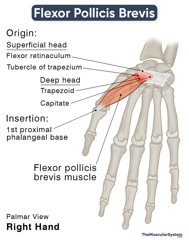
| Origin | Superficial head: Tubercle of the trapezium and the attached flexor retinaculum Deep head: Trapezoid and capitate |
| Insertion | Radial side of the 1st proximal phalangeal base |
Origin
The muscle originates via two heads: superficial and deep. The deep head may be altogether absent at birth in some individuals.
The superficial head originates from the distal side of the tubercle of the trapezium. Some part of it also arises from the flexor retinaculum’s distal border. The deep head originates from the carpal bones, trapezoid, and capitate. The distal carpal row’s palmar ligaments serve as the deep head’s secondary origin.
Insertion
Both heads then travel distally and laterally, narrowing down into a single short tendon to insert into the radial surface of the base of the 1st proximal phalanx. Here, a small sesamoid bone can be found (in 99% of people) embedded in the inserting tendon of the flexor pollicis brevis.
Relations With Surrounding Muscles and Structures
It is the most medial of all 3 thenar muscles, with the opponens pollicis and abductor pollicis brevis lying lateral to it. The superficial head sometimes blends with the opponens pollicis. On the medial side, it has the adductor pollicis muscle next to it.
As it courses distally from the carpal area, its superficial head runs along the inserting tendon of the flexor pollicis longus on its radial side. At the same time, the deep head runs deep to this tendon.
Function
| Action | Thumb flexion at the 1st metacarpophalangeal joint |
As part of the thenar group of muscles, it helps with the flexion of the thumb at the 1st metacarpophalangeal and carpometacarpal joints. It means the muscle helps fold the thumb across the palm and rotate it medially.
These movements help with anything that requires using the thumb with the middle and index fingers, like picking something off the floor or gripping something in your hand.
Innervation
| Nerve | Recurrent branch of the median nerve (C8, T1); Deep branch of ulnar nerve (deep head) |
The nerve supply may vary from one individual to another. Usually, the recurrent branch of the median nerve (C8, T1) innervates both heads. In contrast, the deep branch of the ulnar nerve usually innervates only the deep head. Still, it may also innervate both heads in rare cases.
Superficial palmar branch of radial artery
| Artery | Superficial palmar branch of radial artery |
The primary blood supply comes from the superficial palmar branch of radial artery, while branches of the princeps pollicis and radialis indicis arteries also vascularize this muscle.
References
- Flexor Pollicis Brevis: TeachMeAnatomy.info
- Flexor Pollicis Brevis Muscle: KenHub.com
- Flexor Pollicis Brevis Muscle. Anatomical Study and Clinical Implications: NCBI.NLM.NIH.gov
- Flexor Pollicis Brevis: RAD.Washington.edu
- Determine the Function of the Flexor Pollicis Brevis Muscle: Study.com
- Flexor Pollicis Brevis Muscle: RadioPaedia.org
Della Barnes, an MS Anatomy graduate, blends medical research with accessible writing, simplifying complex anatomy for a better understanding and appreciation of human anatomy.
- Latest Posts by Della Barnes, MS Anatomy
-
Stapedius
- -
Auricular Muscles
- -
Auricularis Posterior
- All Posts


