

Lumbricals of the Hand
By
Della Barnes, an MS Anatomy graduate, blends medical research with accessible writing, simplifying complex anatomy for a better understanding and appreciation of human anatomy.
Last updated:
06/06/2024Della Barnes, MS Anatomy
UX/UI Designer at - AdobeDella Barnes, an MS Anatomy graduate, blends medical research with accessible writing, simplifying complex anatomy for a better understanding and appreciation of human anatomy.
What are the Lumbricals of the Hand
The lumbricals are 4 short intrinsic muscles of the hand, primarily responsible for the movements of the fingers. Their worm-like appearance earns them the name Lumbricals, derived from ‘Lumbricidae’, the Latin name for the earthworm family. The lumbricals belong to the group of short muscles of the hand, along with the dorsal and the palmar interossei.
Anatomy
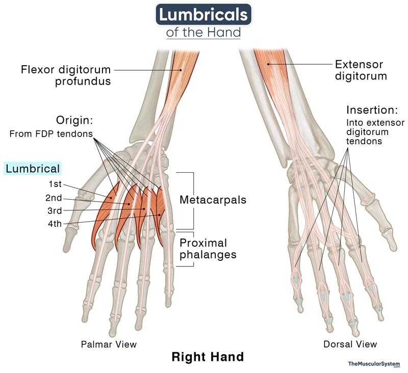
The 4 lumbricals of the hand are numbered from 1st to 4th from lateral to medial side, according to their attachment to the index to little fingers. The 1st and 2nd lumbricals are unipennate muscles, while the 3rd and 4th lumbricals are bipennate.
Location and Attachments
The muscles are located between the 2nd to 5th metacarpal bones, with the palmar fascia lying superficially.
One unique feature of these muscles is that they all originate from and insert into tendons.
| 1st lumbrical | 2nd lumbrical | 3rd lumbrical | 4th lumbrical | |
|---|---|---|---|---|
| Origin | The radial/lateral side and the palmar surface of the flexor digitorum profundus (FDP) tendon of the index finger (2nd digit) | The radial/lateral side and the palmar surface of the FDP tendon of the middle finger (3rd digit) | The ulnar/medial side of the FDP tendon of the middle finger; The radial or lateral side of the FDP tendon of the ring finger (4th digit) | The ulnar/medial side of the FDP tendon of the ring finger; The radial or lateral side of the FDP tendon of the little finger (5th digit) |
| Insertion | The radial margin of the dorsal digital expansion of the extensor digitorum at the 2nd proximal phalanx | The radial margin of the dorsal digital expansion of the extensor digitorum at the 3rd proximal phalanx | The radial margin of the dorsal digital expansion of the extensor digitorum at the 4th proximal phalanx | The radial margin of the dorsal digital expansion of the extensor digitorum at the 5th proximal phalanx |
Origin
As evident from the above table, each lumbrical muscle originates from the tendons of the FDP muscle of its corresponding finger. The FDP inserts into the palmar surfaces of the 2nd to 5th distal phalanges. A point to note here is that the FDP is a partial antagonist of the lumbricals, which means they arise from the tendons of their antagonist.
Insertion
From the point of origin, the narrow muscles course distally and along the lateral margin of the corresponding finger, opposite to its metacarpophalangeal joint. The 1st to 4th lumbricals are then inserted into the tendinous extensor expansion of the extensor digitorum of the 2nd to 5th digit.
Variations
Studies have shown certain variations in their number, form, and attachments. Sometimes, the 1st lumbrical may be divided into two muscles, or the 2nd lumbrical may be bipennate instead of unipennate. The 3rd lumbrical may sometimes be absent altogether. Apart from these, there may be variations in their points of attachment and nerve and arterial supply.
Function
| Action | Finger flexion at the metacarpophalangeal joints; finger extension at the interphalangeal joints |
Finger Flexion and Extension
Since both their attachment points are in tendons, these muscles are highly mobile. Additionally, the muscles cross over from the palmar side towards the dorsal side of the fingers. Their inserting tendons cross the metacarpophalangeal (MCP) joints on the side of the palm and then travel further distally to insert into the fingers’ dorsal side (dorsal digital expansion).
This makes the lumbricals capable of both flexion and extension. They flex the 2nd to 5th MCP joints and extend the proximal (PIP) and distal (DIP) interphalangeal joints of the 2nd to 5th digits. These two actions are responsible for all the complex movements of the fingers and hand and their general dexterity. So, these muscles help with writing, sewing, and gripping something with your finger.
Some of the actions of the lumbricals are antagonistic to those of the FDP muscle, which flexes the PIP and DIP joints.
Proprioception
Proprioception is the body’s ability to know its own location, movement, and any change in position. It enables you to locate and point your right eye with your left index finger, even with your eyes closed, or to know whether you are walking on grass or concrete without looking down.
Studies have suggested the lumbricals’ involvement in proprioception at the interphalangeal joints of the 2nd to 5th digits. These muscles possess the highest number of spindle fibers in the upper extremity despite being among the smallest muscles here. The higher numbers of spindle fibers cause large displacements in the lumbricals, even with the slightest change in flexion at the PIP and DIP joints. As a result, the lumbricals are most likely responsible for the proprioceptive feedback at these joints.
Evolutionary studies have shown an increase in the number of these spindle fibers from non-primate mammals to primates, being the highest in humans. It has led scientists to believe this may be directly connected to whether the forelimb is used for locomotion (non-primate mammals), collecting food, or tool usage (humans).
Innervation
| Nerve | 1st and 2nd: Digital branch of the median nerve (C8-T1) 3rd and 4th: Deep branch of the ulnar nerve (C8-T1) |
A simple mnemonic might help to remember these:
“1 2 me, 3 4 u” (One to me, three for you)
1st and 2nd lumbricals are innervated by the medial nerve (me), while the 3rd and 4th are innervated by the ulnar nerve (u).
Blood Supply
| Artery | Dorsal metacarpal arteries |
The lumbricals primarily receive their arterial supply from the dorsal carpal arch, which is part of the anastomotic network of blood vessels on the back of the hand. Additionally, arteries from the palmar side contribute to their blood supply.
The 1st and 2nd lumbricals are vascularized by the 1st and 2nd dorsal metacarpal arteries, along with the dorsal digital arteries. The 3rd and 4th lumbricals receive blood from the 3rd and 4th dorsal metacarpal arteries, as well as the 2nd and 3rd common palmar digital arteries, which originate from the superficial palmar arch.
References
- Anatomy, Shoulder and Upper Limb, Hand Lumbrical Muscles: NCBI.NLM.NIH.gov
- Lumbricals: WheelessOnline.com
- Lumbrical Muscles of the Hand: KenHub.com
- Lumbricals (Hand): TeachMeAnatomy.info
- Lumbrical Muscles of the Hand: RadioPaedia.org
- Lumbrical Muscles of Hand: IMAIOS.com
- Lumbrical Muscles (Hand, Anatomy): GPNotebook.com
Della Barnes, an MS Anatomy graduate, blends medical research with accessible writing, simplifying complex anatomy for a better understanding and appreciation of human anatomy.
- Latest Posts by Della Barnes, MS Anatomy
-
Tensor Tympani
- -
Stapedius
- -
Auricularis Posterior
- All Posts


