

Tensor Fasciae Latae
By
Della Barnes, an MS Anatomy graduate, blends medical research with accessible writing, simplifying complex anatomy for a better understanding and appreciation of human anatomy.
Last updated:
17/07/2025Della Barnes, MS Anatomy
UX/UI Designer at - AdobeDella Barnes, an MS Anatomy graduate, blends medical research with accessible writing, simplifying complex anatomy for a better understanding and appreciation of human anatomy.
What is the Tensor Fasciae Latae
The tensor fasciae latae (pronounced as ten-sor fas-she-ay lah-tay) or TFL is a small, fusiform, paired muscle located on the lateral side of each thigh. It belongs to the group of gluteal muscles, which form the buttocks. Being a long muscle that extends from the hip to the knee, the tensor fasciae latae plays an important role in flexion, rotation, and abduction at the hip and knee joints.
Anatomy
Location and Attachments
| Origin | Anterior superior iliac spine, and the outer lip of the iliac crest |
| Insertion | The Gerdy’s tubercle via the iliotibial tract |
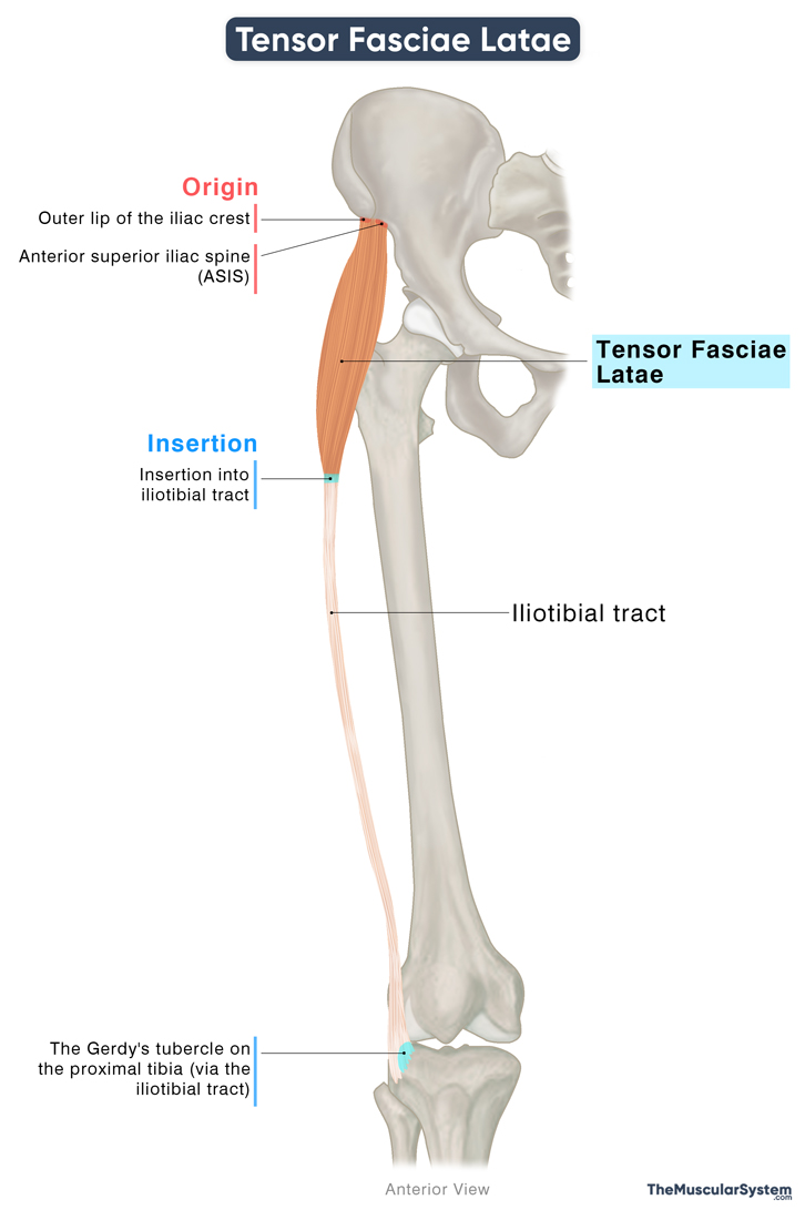
Origin
The muscle originates primarily from the anterior third of the outer lip of the iliac crest and the outer surface of the anterior superior iliac spine (ASIS), which is the prominent bony bump at the front of the pelvis on both sides. Additionally, some fibers arise from the fascia lata, the deep fascia of the thigh that contributes to lower limb stability and movement.
Insertion
The fleshy muscle fibers descend obliquely and quickly converge into a narrow tendon. This tendon merges with the iliotibial tract (IT band), a thick, fibrous band of connective tissue that runs along the lateral aspect of the thigh. The IT band reinforces the fascia lata and continues downward to insert into Gerdy’s tubercle, located on the lateral condyle of the proximal tibia, just below the knee joint.
The iliotibial tract is an extension of the fascia lata, playing a crucial role in stabilizing both the hip and knee joints, particularly during activities such as walking, running, and standing on one leg.
Relations With Surrounding Muscles and Structures
It is a superficial muscle located on the anterolateral side of the thigh, lying just beneath the skin. At its superior end, the TFL is positioned lateral to the hip joint, between the gluteus medius posteriorly and the sartorius anteriorly. In this region, it lies superficial to the gluteus minimus and lateral to the rectus femoris.
As the muscle descends, it courses between the layers of the fascia lata, where it ultimately inserts into the iliotibial tract. Here, it blends with the fibers of the gluteus maximus, which also contribute to the IT.
Function
| Action | Abducting, medially rotating, and flexing the thigh at the hip joint Externally rotating the tibia (lower leg) at the knee joint Stabilizing the hip and knee |
Originating from the ischium and working closely with the iliotibial tract, this muscle helps stabilize both the hip and knee joints, and thus the overall torso. For instance, it prevents the legs from collapsing when you walk, or shift your weight while standing still. It helps with the following actions at these joints:
At the Hip Joint
Hip Abduction
- Works synergistically with the gluteus medius and minimus muscles to lift the leg laterally or to the sides, like during side leg raises.
- Also collaborates with the gluteus maximus via the iliotibial tract to assist in abduction and stabilize the hip.
Medial (Internal) Rotation of the Hip
- Assists gluteus medius and minimus in rotating the thigh inward, like often done when maneuvering a ball during sports like soccer.
Hip Flexion
- Supports the rectus femoris, a muscle of the quadriceps group, in lifting the thigh forward, such as when climbing stairs.
At the Knee Joint
The tensor fasciae latae influences movement at the lower leg through the iliotibial tract, which attaches to the Gerdy tubercle on the tibia — the larger of the two lower leg bones.
Lateral Rotation of the Tibia (Lower Leg)
- Indirectly helps with lateral rotation of the tibia, which means twisting your lower leg outward while the upper leg stays stable, like during dynamic movements, such as dancing or playing soccer
- Helping with flexing or bending the knee, but only when the knee is already bent at least 30°. For example, when you are trying to sit in a chair, the knees need to bend past 30°, so the TFL helps.
Antagonists
Muscles that perform opposite actions are considered antagonists. The TFL assists with hip abduction, medial rotation, and flexion. Therefore, muscles that produce the opposite movements, such as the hip extensors (e.g., gluteus maximus and hamstrings), hip adductors (e.g., adductor longus, brevis, and magnus), and lateral rotators (e.g., piriformis, obturator internus, superior and inferior gemelli), can be considered antagonistic to the TFL.
Innervation
| Nerve | Superior gluteal nerve (L4-S1) |
The muscle receives its blood supply from the superior gluteal nerve, arising from the posterior divisions of the 4th-5th lumbar, and 1st sacral nerve (L4, L5, and S1) roots of the sacral plexus.
Blood Supply
| Artery | Lateral circumflex femoral artery and superior gluteal artery |
The primary blood supply to the TFL muscle comes from the ascending branch of the lateral circumflex femoral artery and the deep branch of the superior gluteal artery.
References
- Tensor Fascia Lata:TeachMeAnatomy.info
- Anatomy, Bony Pelvis and Lower Limb: Tensor Fasciae Latae Muscle: NCBI.NLM.NIH.gov
- Tensor Fascia Lata Muscle: Kenhub.com
- Tensor Fascia Lata: Rad.Washington.edu
- Tensor Fascia Lata: Elsevier.com
- Tensor Fascia Lata: GetBodySmart.com
- Tensor Fascia Lata: IMAIOS.com
Della Barnes, an MS Anatomy graduate, blends medical research with accessible writing, simplifying complex anatomy for a better understanding and appreciation of human anatomy.
- Latest Posts by Della Barnes, MS Anatomy
-
Tensor Tympani
- -
Stapedius
- -
Auricularis Posterior
- All Posts


