Transverse Perineal
Last updated:
19/06/2025Della Barnes, an MS Anatomy graduate, blends medical research with accessible writing, simplifying complex anatomy for a better understanding and appreciation of human anatomy.
What is the Transverse Perineal
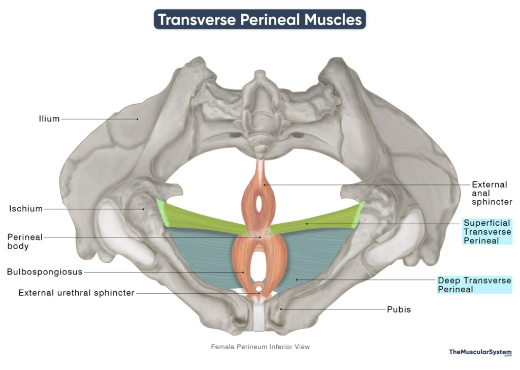
The transverse perineal muscles, collectively known as the transversus perinei, consist of two paired muscles: the superficial transverse perineal and the deep transverse perineal muscles. These slender muscle bands play a crucial role in maintaining the stability and function of the perineum, the region between the thighs that encompasses the genital and anal areas.
Anatomy
Location and Attachments
Although typically associated with one another, these muscles are anatomically separate. The superficial transverse perineal muscle is located within the superficial perineal pouch. In contrast, the deep transverse perineal muscle resides in the deep perineal pouch.
| Muscle | Origin | Insertion |
| Superficial Transverse Perineal | Front of the ischial tuberosity | Fibers of the contralateral STP muscle (encircling perineal body) |
| Deep Transverse Perineal | Inferior ischial ramus | Fibers of the contralateral DTP muscle (encircling perineal body) |
Origin
Both the muscles originate from the ischium, though at slightly different spots.
The superficial transverse perineal muscle arises via small tendons from the inner and frontal part of the ischial tuberosity.
In contrast, the deep transverse perineal muscle originates from the inferior ischial ramus, which lies just in front and slightly below the origin of the superficial muscle.
Insertion
From their points of origin, both muscles course medially, or transversely, toward the perineal body. It is a small, fibromuscular mass located in the center of the perineum, between the vagina and the anus in females, and between the scrotum and the anus in males. It serves as a key attachment point for several pelvic floor muscles, helping to support the pelvic organs.
For a long time, both muscles were believed to insert entirely into the perineal body. However, recent studies suggest that most of the muscle fibers actually pass around the perineal body to join the fibers coming from the opposite side (contralateral muscle). This creates a continuous, intertwined sling-like structure just in front of the anus. A few muscle fibers may also insert directly into the perineal body.
Specifically, fibers from the superficial transverse perineal blend with those of the contralateral superficial muscle, and the deep transverse perineal muscle likewise converges with its counterpart on the opposite side.
Relations With Surrounding Muscles and Structures
Superficial Transverse Perineal
The intertwining fibers of the transverse perineal muscles meet and overlap fibers from other perineal muscles in the perineal body. The point where the fibers from the contralateral superficial transverse perineal muscles meet has the external anal sphincter muscle lying in the back, while the bulbospongiosus muscle lies in front.
In fact, in some anatomical variations, the fibers of superficial transverse perineal muscle might even blend with those of the external anal sphincter and/or bulbospongiosus.
Deep Transverse Perineal
It is located at the same level as the external urethral sphincter in the pelvis, within the deep perineal pouch. In females, the vagina passes between the left and right deep transverse perineal muscles.
Function
| Action | Providing structural support to the perineum |
Since both muscles contribute to the tendinous raphe of the perineal body, their primary function involves stabilizing the perineum and supporting the pelvic and perineal organs and structures.
In women, these muscles assist in resisting increases in intra-abdominal pressure, help prevent pelvic organ prolapse, and contribute significantly to the recovery and restoration of pelvic floor function after childbirth.
Innervation
| Nerve | Perineal branch of the pudendal nerve (S2-S4) |
The perineal nerve, a terminal branch of the pudendal nerve originating from the 2nd to 4th sacral nerve roots (S2 to S4), innervates the transverse perineal muscles.
Blood Supply
| Artery | Perineal artery |
Blood supply to these muscles comes from the perineal artery, a branch of the internal pudendal artery.
References:
- Superficial Transverse Perineal Muscle: Radiopaedia.org
- Deep Transverse Perineal Muscle: Radiopaedia.org
- Anatomy, Abdomen and Pelvis: Superficial Perineal Space: NCBI.NLM.NIH.gov
- Perineal Region: Kenhub.com
- Deep Transverse Perinei Muscle: GPNotebook.com
- The Perineum: TeachMeAnatomy.info
Della Barnes, an MS Anatomy graduate, blends medical research with accessible writing, simplifying complex anatomy for a better understanding and appreciation of human anatomy.
- Latest Posts by Della Barnes, MS Anatomy
-
Tensor Tympani
- -
Stapedius
- -
Auricularis Posterior
- All Posts


