

External Urethral Sphincter
By
Della Barnes, an MS Anatomy graduate, blends medical research with accessible writing, simplifying complex anatomy for a better understanding and appreciation of human anatomy.
Last updated:
23/06/2025Della Barnes, MS Anatomy
UX/UI Designer at - AdobeDella Barnes, an MS Anatomy graduate, blends medical research with accessible writing, simplifying complex anatomy for a better understanding and appreciation of human anatomy.
What is the External Urethral Sphincter
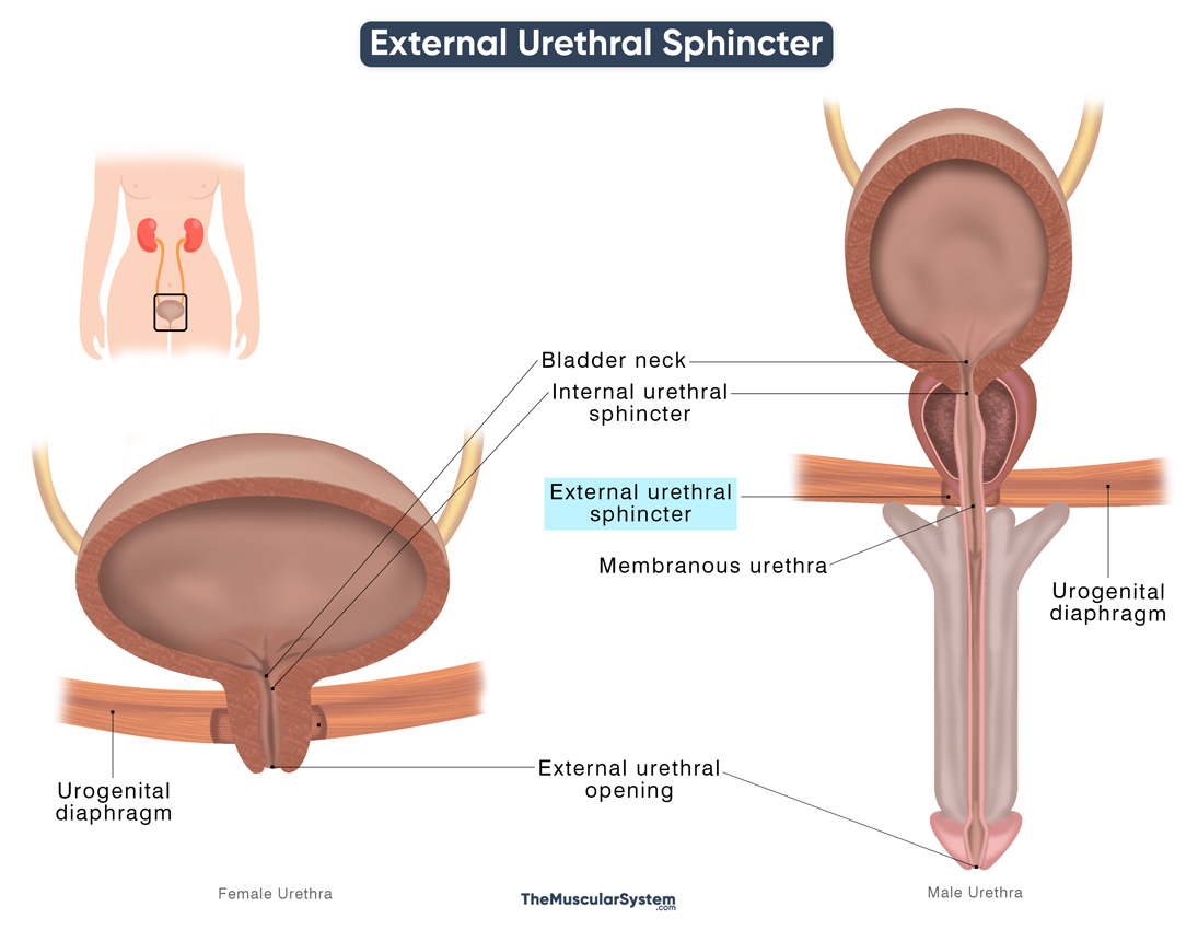
The external urethral sphincter is one of the two urethral sphincter muscles that play a vital role in controlling the flow of urine. It is a voluntary skeletal muscle, meaning it is controlled by the somatic nervous system and can be consciously controlled. The other urethral sphincter, the internal urethral sphincter, is made up of smooth muscle, and thus it is an involuntary muscle.
The muscle belongs to the urogenital triangle, more specifically to the deep perineal pouch. It surrounds the membranous part of the urethra, helping with controlling its opening and closing.
Anatomy
The origin and insertion of the muscle remain the same for both sexes. However, its path and structure vary, being much more complex in females compared to males.
Location and Attachments
| Origin | The ischial rami, inferior pubic rami, and the attached fascia |
| Insertion | Fibers from the contralateral side of the muscle |
Male External Urethral Sphincter
Origin
The muscle fibers arise from both sides of the pelvis, at the junction where the ischial ramus meets the inferior pubic ramus. Some fibers also originate from the adjacent fascia. The point of origin may extend over approximately 2 cm of the bony surface.
Structure and Insertion
From their origin, the muscle fibers loop around the urethra. At the back of the urethra, the left and right external urethral sphincter fibers meet and fuse, contributing to a fibrous seam that further supports the muscle’s function.
The innermost fibers of the muscle form a muscular ring that surrounds the middle part of the urethra, also known as the membranous urethra.
Female External Urethral Sphincter
Origin
The origin of the muscle is the same in females as in males.
Structure and Insertion
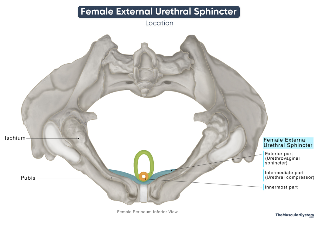
The female external urethral sphincter can be divided into 3 parts:
- The first, or innermost part is composed of circular fibers that surround the proximal part of the female urethra, forming a true muscular ring or “sphincter” around it. The muscular fibers forming the front part of the ring are thicker than those at the back.
- The second, or intermediate part, known as the urethral compressor muscle, has attachments at the ischial and pubic ramus. The muscle fibers from each side course around the front of the urethra to fuse with the fibers from the opposite side (contralateral fibers). As a result, they form a loop around the urethra.
- The third, or most exterior part, known as the urethrovaginal sphincter, is a large, flat fibrous band that courses posteriorly from its origin at the ischial and pubic ramus to go around both the urethra and vagina, extending to the vestibular bulbs.
Relations With Surrounding Muscles and Structures
The external urethral sphincter is located around the membranous urethra, making it distal to the internal urethral sphincter which lies at the vesicourethral junction (the point where the bladder neck meets the urethra). Although traditionally described as separate muscles — one smooth (internal) and one skeletal (external) — modern anatomical studies suggest that the internal and external urethral sphincters may form a continuous, functionally integrated layer known as the urethral sphincter complex that surrounds the mid-portion of the urethra.
In both sexes, the muscle lies in front of the perineal body and is superior to the perineal membrane. In males, it is positioned just beneath the prostate. In females, it lies directly below the bladder, since there is no prostate present.
The bulbourethral glands, a pair of small glands in the male reproductive system, are located near the external urethral sphincter. These glands are often partially embedded within the muscle fibers of the sphincter.
Function
| Action | Allowing voluntary control over the urethral opening, and maintaining urinary continence |
The muscle’s functions are interrelated with those of the internal urethral sphincter, as the two work together to control the opening of the urethra. When the bladder is full, it stretches and sends signals to the nervous system, triggering the smooth muscles of the bladder (the detrusor muscle) to contract and expel urine. The internal urethral sphincter, composed of smooth muscle and controlled by the autonomic nervous system, also relaxes and opens.
In contrast, the external urethral sphincter is a voluntary skeletal muscle, meaning it can be consciously kept closed to delay urination until an appropriate time. This voluntary control is coordinated by motor neurons located in Onuf’s nucleus of the sacral spinal cord (S2–S4).
So, the primary function of this muscle is to prevent urine flow unless it is voluntarily relaxed. This makes it a critical structure for maintaining urinary continence.
In females, the muscle helps constrict the vaginal opening as well as the urethra.
Innervation
| Nerve | Perineal branch of the pudendal nerve (S2-S4) |
External Urethral Sphincter
Like most of the muscles in the perineal region, the primary innervation to the muscle comes from the pudendal nerve’s perineal branch. The pudendal nerve arises from the ventral rami of the 2nd to 4th sacral spinal nerves (S2 to S4), with motor fibers originating specifically from Onuf’s nucleus.
Blood Supply
| Artery | In males: Bulbourethral artery In female: Internal pudendal artery |
In males, the muscle receives its primary blood supply from the bulbourethral artery, which branches from the internal pudendal artery, and also supplies the bulb of the penis.
In females, the main blood supply comes from the internal pudendal artery, which is a branch of the internal iliac artery.
References:
- Anatomy, Abdomen and Pelvis, Sphincter Urethrae: NCBI.NLM.NIH.gov
- Urethral Sphincters: Kenhub.com
- Male External Urethral Sphincter (Right Half): Elsevier.com
- Female External Urethral Sphincter: Elsevier.com
- Male External Urethral Sphincter: IMAIOS.com
- Female External Urethral Sphincter: IMAIOS.com
- Urethra: ClevelandClinic.org
Della Barnes, an MS Anatomy graduate, blends medical research with accessible writing, simplifying complex anatomy for a better understanding and appreciation of human anatomy.
- Latest Posts by Della Barnes, MS Anatomy
-
Tensor Tympani
- -
Stapedius
- -
Auricularis Posterior
- All Posts


