

Levator Ani
By
Della Barnes, an MS Anatomy graduate, blends medical research with accessible writing, simplifying complex anatomy for a better understanding and appreciation of human anatomy.
Last updated:
02/02/2026Della Barnes, MS Anatomy
UX/UI Designer at - AdobeDella Barnes, an MS Anatomy graduate, blends medical research with accessible writing, simplifying complex anatomy for a better understanding and appreciation of human anatomy.
What is the Levator Ani
The levator ani is a funnel-shaped muscle that consists of three thin muscles, the Iliococcygeus, pubococcygeus, and puborectalis. It is the largest of the two muscles forming the pelvic floor, with the other one being the coccygeus. So its primary functions are to support the pelvic viscera, stabilize the pelvis, and help maintain continence.
Anatomy
The three smaller muscles forming the levator ani cover most of the pelvic floor. In order of their point of origin from back to front, these muscles are the (i) iliococcygeus, (ii) pubococcygeus, and (iii) puborectalis.
Location and Attachments
| Component | Origin | Insertion |
| Iliococcygeus | Inner surface of ischium and the back of the obturator fascia | Coccyx and anococcygeal raphe |
| Pubococcygeus | Inferior rami of pubis, and the front of the obturator fascia | Coccyx and anococcygeal raphe |
| Puborectalis | Lower part of the pubic symphysis, and the superior fascia of the urogenital diaphragm | Anococcygeal raphe |
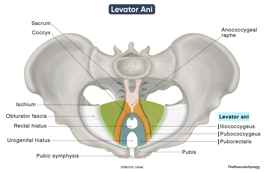
The three muscles originate one by one from the back of the pelvis and travel around the rectum to form a funnel or v-shaped structure.
Iliococcygeus
The iliococcygeus, the posterior-most muscle in the levator ani group, originates from the inner front surface of the ischium bone, and the inner back surface of the obturator fascia — the tendinous fascia of the obturator muscle in the hip.
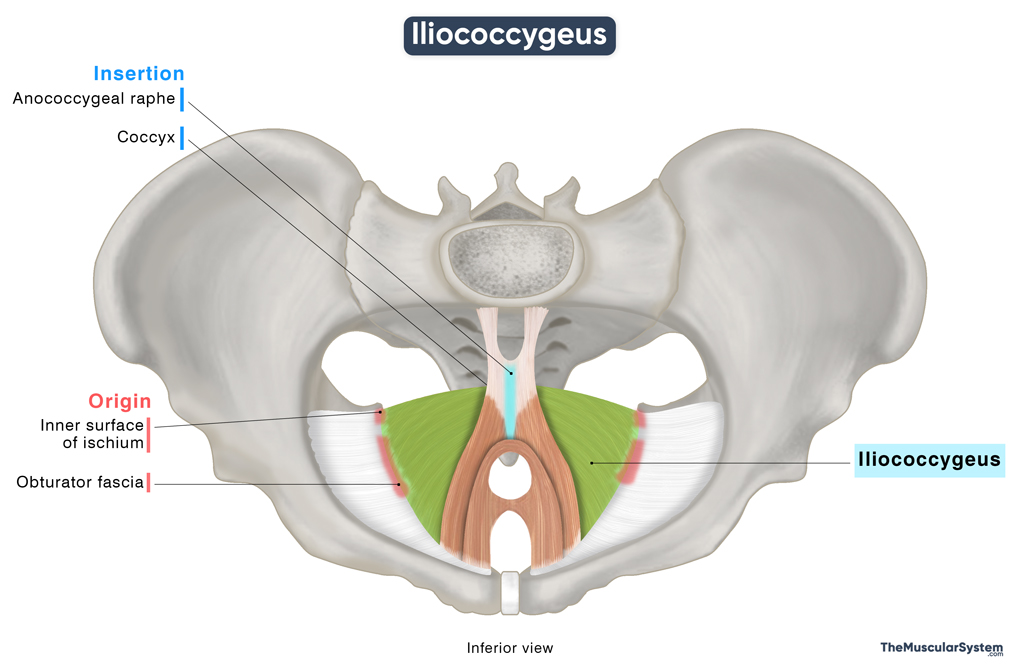
From there, the muscle fibers course downward towards the back to insert into the coccyx. These are the most laterally running fibers of the levator ani. Some part of the muscle travels slightly anteriorly to blend with the pubococcygeus and contribute to the anococcygeal raphe or anococcygeal ligament. It is a fibrous ridge along the midline of the body that connects the coccyx and the anorectal junction.
Pubococcygeus
The pubococcygeus is the next thin muscle in the funnel-like structure, arising from the inner surface of the pubis bone, and the front part of the obturator fascia. The muscle fibers then travel almost horizontally towards the back on both sides around the rectum.
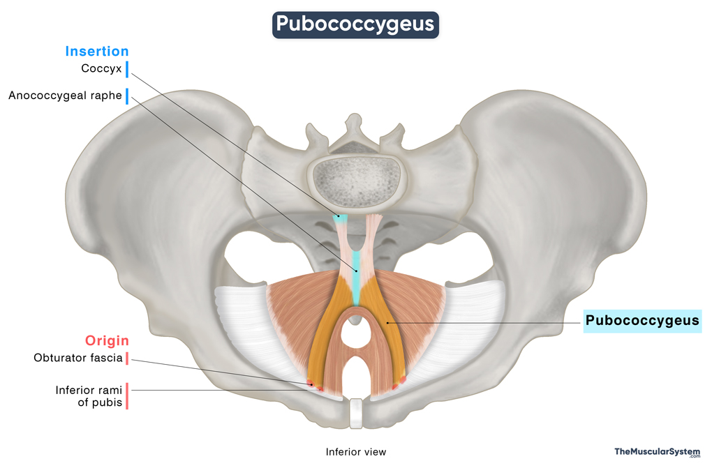
When the fibers reach the back, some part of it inserts into the lower part of the coccyx. The rest of the fibers from the pubococcygeus muscles, coming from the opposite sides, come together to form the anococcygeal raphe.
The pubococcygeus muscle varies slightly between sexes. In males, some fibers form the puboprostaticus, while in females, they form the pubovaginalis. These are functional subdivisions rather than entirely separate muscles.
Puborectalis
The puborectalis, also referred to as the puboanalis, is the third and anterior-most muscle of the levator ani. It arises from the posterior surface of the body of the pubis, on each side of the pubic symphysis. Some fibers also rise from the upper part of the urogenital diaphragm’s fascia.
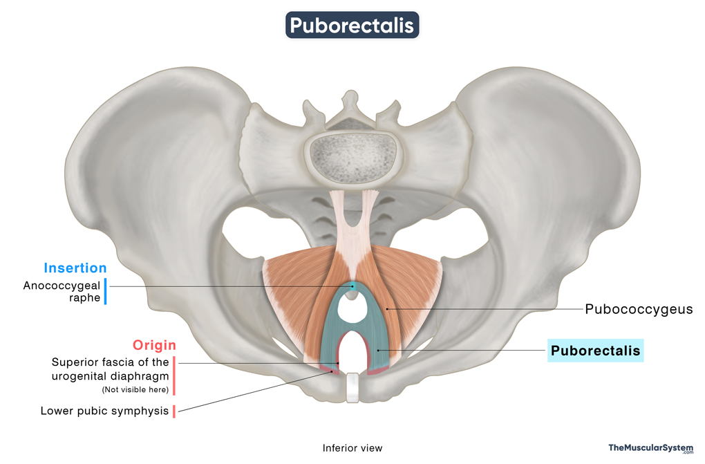
Unlike most muscles with well-defined insertion points, the puborectalis loops around the anorectal junction to form a U-shaped sling. It originates from the pubis on one side, loops around the lower end of the rectum, and inserts into the pubis on the opposite side.
Relations With Surrounding Muscles and Structures
The levator ani is an essential component of the pelvic floor. Superior to this muscle group lie the pelvic viscera, including the bladder, rectum, and prostate in males, or the uterus and vagina in females. Inferiorly, it forms the roof of the perineum. Anteriorly, it is related to the pubic symphysis, and posteriorly to the coccyx.
As the puborectalis loops around the urethra, vagina (when present), and rectum, it forms a U-shaped gap along the midline of the pelvic floor known as the levator hiatus, through which the urethra, rectum, and vagina pass. This hiatus is bordered by the other two levator ani muscles and is an important anatomical landmark and a common site of pelvic organ prolapse, particularly in women.
Function
| Action | Stabilizing the pelvis and supporting the pelvic viscera Helping to resist increased intra-abdominal pressure Controlling the rectum and urethra |
As a whole, the muscle group performs the following functions:
- Supporting the pelvic viscera, which means holding organs like the bladder, rectum, and uterus in their place and preventing prolapse through the levator hiatus.
- Assisting with postural stability and functional movements such as standing, walking, and bending by maintaining the integrity of the pelvic floor.
- Contributing to sexual function by supporting vaginal and urethral tone, aiding orgasm in females, and helping maintain erections and support ejaculation in males.
Each of the three muscles in this group also plays a distinct role in supporting the overall function of the pelvic floor.
Functions of the Iliococcygeus Muscle
It supports the pelvic floor and contributes to the anococcygeal raphe, reinforcing it as a midline structure that anchors surrounding pelvic fascia and muscles.
Functions of the Pubococcygeus Muscle
It plays a key role in urinary control by supporting the urethra and helping to close the urogenital hiatus. It also assists in managing intra-abdominal pressure during activities such as coughing or sneezing. This is why people with a weak pubococcygeus, such as older people, pregnant women, or women who have given birth, are more at risk of experiencing urinary incontinence, which means difficulty holding urine in. Its extensions have their own role:
- Puboprostaticus (in men): Supports the prostate and aids in ejaculation.
- Pubovaginalis (in women): Supports the vagina, and assists in positioning the baby’s head during childbirth.
Functions of the Puborectalis Muscle
The puborectalis, the smallest of all the levator ani muscles, works with the external anal sphincter and internal anal sphincter, to control bowel movements. Thanks to its U-shaped loop around the anorectal junction, the puborectalis muscle helps maintain continence by keeping the rectum closed. When the muscle relaxes, it opens the rectum, allowing defecation. The muscle also helps with the closure of the urethra.
In animals like dogs and horses, the levator ani muscle helps move the tail, especially during actions like wagging.
Antagonists
Since the levator ani’s actions primarily involve supportive and sphincteric control of the levator hiatus, rather than moving a joint through a wide range of motion, it does not have any antagonists in the traditional sense.
Innervation
| Nerve | S3-S4, and pudendal nerves |
The pelvic or superior surface of the levator ani group is innervated by branches of the 3rd and 4th sacral nerves. The perineal or inferior surface may also receive additional innervation from the pudendal nerve, specifically through its perineal and/or inferior rectal branches.
Blood Supply
| Artery | Inferior gluteal, internal pudendal, and inferior vesical arteries |
These muscles receive their blood supply from several branches of the internal iliac artery, including the inferior gluteal artery, inferior vesical artery, and the internal pudendal artery.
References:
- Anatomy, Abdomen and Pelvis: Levator Ani Muscle: NCBI.NLM.NIH.gov
- Levator Ani Muscle: Kenhub.com
- Levator Ani Muscle | Structure, Innervation & Function: Study.com
- Levator Ani: IMAIOS.com
- Iliococcygeus Muscle: Elsevier.com
- Pubococcygeus: TeachMeAnatomy.info
- Puborectalis: TeachMeAnatomy.info
- The Pelvic Floor: TeachMeAnatomy.info
- Levator Ani Muscle: Radiopaedia.org
Della Barnes, an MS Anatomy graduate, blends medical research with accessible writing, simplifying complex anatomy for a better understanding and appreciation of human anatomy.
- Latest Posts by Della Barnes, MS Anatomy
-
Tensor Tympani
- -
Stapedius
- -
Auricularis Posterior
- All Posts


