

Psoas Minor
By
Della Barnes, an MS Anatomy graduate, blends medical research with accessible writing, simplifying complex anatomy for a better understanding and appreciation of human anatomy.
Last updated:
14/05/2025Della Barnes, MS Anatomy
UX/UI Designer at - AdobeDella Barnes, an MS Anatomy graduate, blends medical research with accessible writing, simplifying complex anatomy for a better understanding and appreciation of human anatomy.
What is the Psoas Minor
The psoas minor is a thin, paired, fusiform muscle in the posterior abdomen and pelvic region. It is present in only about 40-60% of the population and may appear on one or both sides. When both muscles are present, they may differ in length between the left and right sides.
Functionally, the psoas minor serves as a weak flexor of the lumbar spine. In individuals where it is absent, this action is performed solely by the psoas major.
Anatomy
Location and Attachments
| Origin | T12 and L1 vertebral bodies and the adjacent intervertebral disk |
| Insertion | Iliopubic eminence and pectineal line |
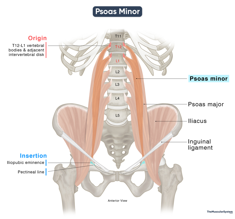
Origin
The muscle originates from the lateral surface of the 12th thoracic and 1st lumbar vertebral bodies, and the intervertebral disk between them.
Insertion
From its point of origin, the psoas minor muscle descends along the medial border of the psoas major. As it courses downward, the muscle fibers gradually taper and flatten, forming a ribbon-like belly that quickly transitions into a long, slender tendon. This tendon continues its path to insert onto the pectineal line of the pubis (also known as the pecten pubis) and the iliopectineal eminence—a bony prominence marking the junction of the ilium and pubis bones.
Though it is located close to the psoas major muscle, the psoas minor muscle does not go past the inguinal ligament on the lower end. Also, despite its name and location, it is not part of the iliopsoas muscle, which consists only of the psoas major and iliacus.
Relations With Surrounding Muscles and Structures
Being a small muscle that fits entirely into the anterior and medial side of the psoas major, the two muscles share the same relations with many surrounding organs and structures. On both sides of the body, the kidneys and ureters lie laterally and in front of the muscle. On the right side, the inferior vena cava and the terminal portion of the ileum (the lowest part of the small intestine) are situated anterior to the psoas minor. Meanwhile, on the left side, the descending colon passes the front of the muscle.
Function
| Action | Flexing the lumbar vertebral columns |
Although this thin muscle is too weak to play a major role in movement or stability, when present, it assists the psoas major in flexing the lumbar vertebral column to bend the trunk forward or lift it from a supine position, such as during a sit-up. However, its absence has no impact on the body’s overall functionality.
Antagonists
Although it’s not as powerful as the other muscles in the region, its role in trunk flexion can be seen as antagonistic to the erector spinae muscle group, which is responsible for extending the trunk.
Innervation
| Nerve | Anterior ramus of L1 spinal nerve |
The muscle receives innervation from the anterior ramus of the 1st lumbar spinal nerve (L1), which is a branch of the lumbar plexus.
Blood Supply
| Artery | Lumbar arteries |
The lumbar arteries provide the primary vascular supply to the muscle. Additional blood supply comes from the iliolumbar and obturator arteries, which branch from the internal iliac artery.
References:
- Psoas Minor Muscle: Kenhub.com
- Psoas Minor: TeachMeAnatomy.info
- Psoas Minor: Elsevier.com
- Psoas Minor Muscle: Radiopaedia.org
- Psoas Minor: IMAIOS.com
Della Barnes, an MS Anatomy graduate, blends medical research with accessible writing, simplifying complex anatomy for a better understanding and appreciation of human anatomy.
- Latest Posts by Della Barnes, MS Anatomy
-
Tensor Tympani
- -
Stapedius
- -
Auricularis Posterior
- All Posts


