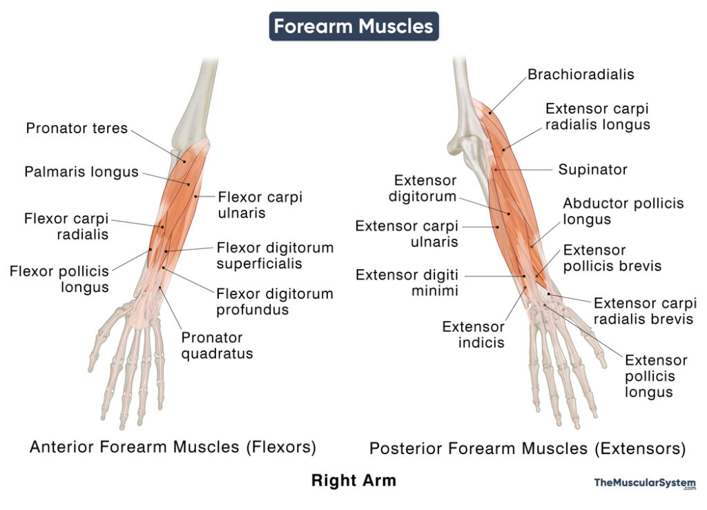
Forearm Muscles
The part of the human arm between the elbow and the wrist is commonly called the forearm. The anatomical term for the forearm is the antebrachium. Two long bones, the radius and ulna, structure this section of the arm, also acting as the point of attachment for several muscles originating in this area.
The forearm muscles are divided into two compartments based on location and action: the anterior or flexor compartment and the posterior or extensor compartment. There are a total of 19 muscles in the forearm that help move not only the elbow and wrist joints but also the joints in the hand and fingers.
Basic Structure and Names of Muscles in the Forearm
The Anterior (Flexor) Compartment of the Forearm
The anterior compartment contains all the flexors of the forearm, hand, and fingers. These are further divided into three groups: superficial, intermediate, and deep flexors.
Superficial Flexors
All these 4 muscles originate partially or completely via the common flexor tendon from the medial epicondyle of the humerus.
Intermediate Flexors
The FDS can sometimes be included in the superficial extensors of the forearm. But it typically lies in a layer between the superficial and deep muscles, so it is classified into its own ‘intermediate’ layer.
Deep Flexors
The Posterior (Extensor) Compartment of the Forearm
Similar to how the anterior compartment contains all the flexors, all the muscles in the posterior compartment work as the extensors of the forearm, wrist, and hand. They are also grouped as superficial and deep posterior forearm muscles.
There are 11 muscles in total in the extensor compartment.
Superficial Extensors
- Extensor digitorum
- Extensor digiti minimi
- Extensor carpi ulnaris
- Brachioradialis
- Extensor carpi radialis longus
- Extensor carpi radialis brevis
The brachioradialis and the extensor carpi radialis longus and brevis are located laterally, on the radial side of the forearm. Together these 3 muscles are referred to as the mobile wad of Henry, or simply, the mobile wad.
Deep Extensors
- Supinator
- Extensor indicis
- Abductor pollicis longus
- Extensor pollicis brevis
- Extensor pollicis longus
The inserting tendons of the 3 deepest muscles, the abductor pollicis longus, extensor pollicis brevis, and extensor pollicis longus, form the medial and lateral borders of the anatomical snuffbox — the triangular depression on the radial side of the dorsum, at the base of the thumb. These 3 muscles can together be referred to as the anatomical snuffbox muscles.
Functions of the Muscles of the Forearm
As mentioned above, the muscles in the anterior or flexor compartment all work together to flex the forearm and hand, which involves bending the forearm downward at the wrist and folding the fingers. Similarly, the posterior or flexor muscles all work to extend the forearm and hand, that is, extending the wrist upward (like when you do a stop sign) and opening up the fingers.
The intrinsic muscles of the forearm, the ones that insert into the radius or ulna, work to pronate and supinate the forearm and hand. The extrinsic muscles, the ones with their insertion points in the hand bones (carpals, metacarpals, and phalanges) are mostly responsible for flexing and extending the fingers and wrist. They also help with gripping something with the fingers and letting something go.
These actions allow all sorts of arm and hand movements that let us do everything from driving, brushing our teeth, holding a phone, and shaking hands.
References
- Forearm Muscles: What to Know: WebMD.com
- Anatomy, Shoulder and Upper Limb, Forearm Muscles: NCBI.NLM.NIH.gov
- Forearm muscles: GetBodySmart.com
- Forearm Muscles: Anatomy and Movement: Study.com
- Muscles of the Forearm: Osmosis.org
- Muscles in the Anterior Compartment of the Forearm: TeachMeAnatomy.info


