

Vastus Medialis
By
Della Barnes, an MS Anatomy graduate, blends medical research with accessible writing, simplifying complex anatomy for a better understanding and appreciation of human anatomy.
Last updated:
19/08/2025Della Barnes, MS Anatomy
UX/UI Designer at - AdobeDella Barnes, an MS Anatomy graduate, blends medical research with accessible writing, simplifying complex anatomy for a better understanding and appreciation of human anatomy.
What is the Vastus Medialis
The vastus medialis, also known as the vastus internus, is a unipennate muscle located on the inner (medial) side of the thigh. It is part of the anterior compartment of the thigh, and is the smallest of the four quadriceps femoris muscles, which serve as the primary extensors of the knee. The other three muscles in the group are the rectus femoris, vastus lateralis, and vastus intermedius.
Despite being relatively small, vastus medialis has a thick, fleshy belly with a distinctive teardrop shape, earning it the nickname “teardrop muscle.”
Anatomy
Location and Attachments
| Origin | The medial side of the proximal femur, including the intertrochanteric line, pectineal line, and medial lip of the linea aspera |
| Insertion | The base of the patella (kneecap) via the quadriceps femoris tendon The tibial tuberosity via the patellar ligament |
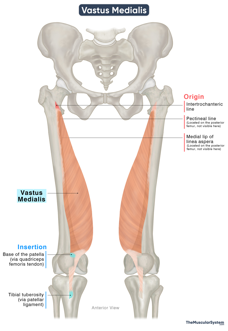
Origin
The muscle has a broad point of origin on the medial surface of the femur, with several points pf origin:
- From the lower part of the intertrochanteric line, the bony ridge below the femoral neck
- From the medial or inner lip of the linea aspera
- The upper part of the medial supracondylar line
- Along the pectineal line
In addition to its attachments to the femur, some part of the muscle also originates from:
- The originating tendons of the adductor magnus
- The upper part of the tendons of the adductor longus
- The adjacent medial intermuscular septum
Structure and Orientation
Being a unipennate muscle, its fibers are aligned obliquely on one side of the central tendon.
Some sources distinguish two parts of the vastus medialis based on the orientation of its muscle fibers. The more vertically aligned fibers that run along the muscle’s long axis form the vastus medialis longus (VML).
As the muscle descends obliquely through the thigh, its fibers spiral slightly around the long axis of the muscle. Near the distal end of the muscle belly, the fibers run more obliquely, lying almost horizontally. This part is known as the vastus medialis obliquus (VMO) and appears as a muscular bulge just above the medial border of the patella.
Though the status of the VML and VMO as anatomically separate parts remains a subject of debate, this difference in fiber orientation is clinically and functionally important.
Insertion
Near the distal end of the femur, the muscle belly narrows into a tendon, which merges with the tendons of the other quadriceps muscles to form the quadriceps femoris tendon. This broad tendon inserts into the base, or superior surface, of the patella.
A portion of this tendon continues from the apex of the patella as the patellar ligament, which inserts into the tibial tuberosity. Through this connection, the vastus medialis, along with the rest of the quadriceps group, is anchored to the tibia and able to act on the lower leg.
Relations With Surrounding Muscles and Structures
As the name implies, the vastus medialis is the most medially positioned muscle of the quadriceps femoris group. Laterally, it is bordered by the vastus intermedius and rectus femoris muscles. The rectus femoris also lies superficial to it, partially covering its superior and lateral aspects.
The sartorius muscle lies superficially on the medial side, while posteriorly, it is separated from the adductor longus and adductor magnus muscles by the medial intermuscular septum.
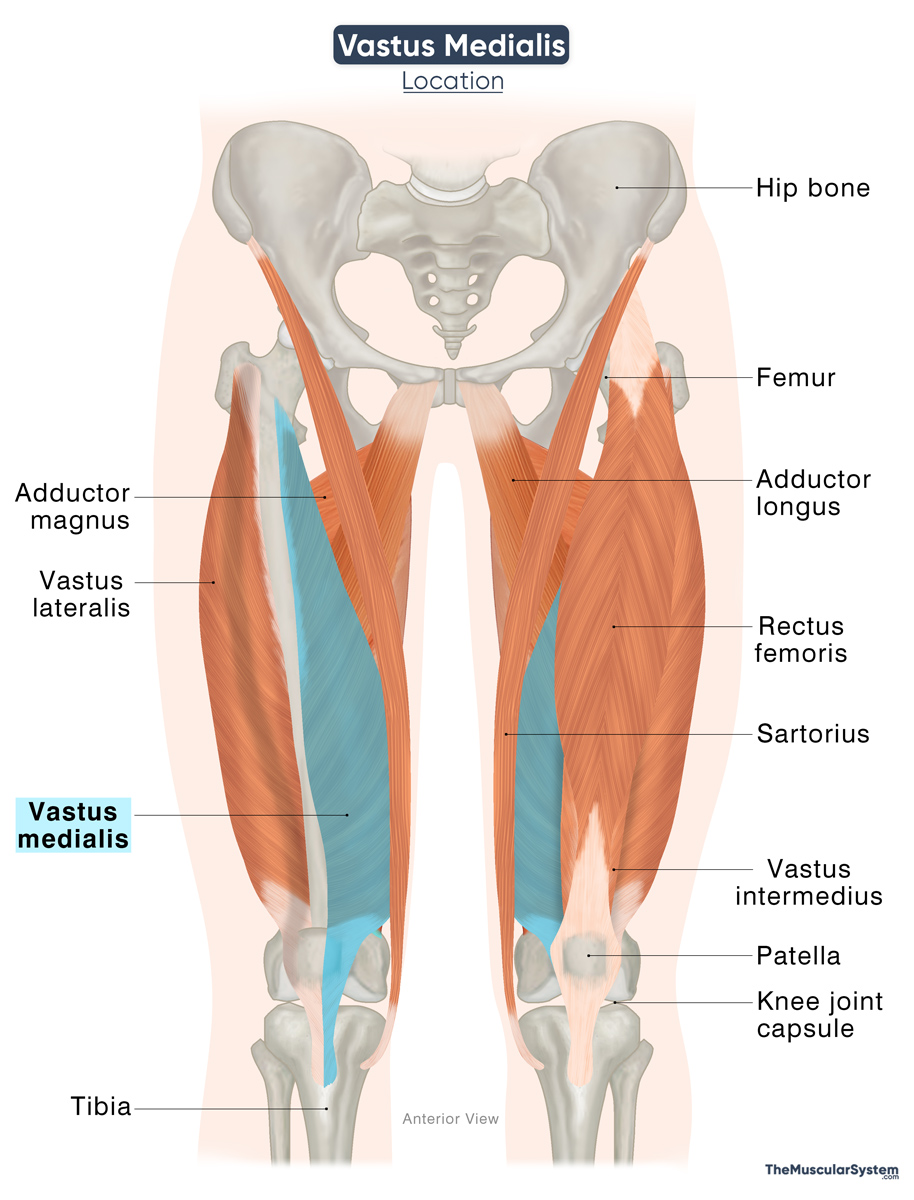
In the mid-thigh region, the muscle contributes to the formation of the adductor canal (Hunter’s canal), an aponeurotic tunnel that allows passage to branches of the femoral nerve and vessels. The vastus medialis forms the anterior wall of this canal, the sartorius forms the medial wall, and the adductor longus and magnus constitute the posterior wall.
Function
| Action | Extending the knee, and stabilizing the knee joint |
With its attachments at the proximal femur, patella, and tibia, the muscle acts on the knee joint, participating in the following functions:
Extension: As part of the quadriceps, it is one of the primary muscles that extend the lower leg at the knee joint. So, the muscle is active during activities like walking, running, climbing, etc.
Stabilization: Its small size and limited attachment to the patella reduce its ability to directly counteract the stronger lateral pull of the vastus lateralis. However, its tendinous fibers contribute indirectly by pulling on the aponeurosis of the vastus intermedius. This combined action helps generate a medial force on the patella that balances the lateral pull, thereby keeping it centered and stabilizing the knee joint.
Antagonists
The hamstrings (biceps femoris, semitendinosus, and semimembranosus) are the primary knee flexors, making them antagonistic to this muscle’s knee-extending action.
Innervation
| Nerve | Femoral nerve (L2-L4) |
The muscle is innervated by the femoral nerve, which arises from the L2 to L4 nerve roots and is the largest branch of the lumbar plexus.
Blood Supply
| Artery | Femoral artery |
The muscle receives its primary blood supply from branches of the femoral artery. Additional blood supply comes from the deep femoral and the genicular arteries.
References
- Vastus Medialis Muscle: Elsevier.com
- Anatomy, Bony Pelvis and Lower Limb: Thigh Quadriceps Muscle: NCBI.NLM.NIH.gov
- Vastus Medialis Muscle: Radiopaedia.org
- Quad Muscles: Clevelandclinic.org
- Vastus Medialis Muscle: GetBodySmart.com
- Vastus Medialis: TeachMeAnatomy.info
Della Barnes, an MS Anatomy graduate, blends medical research with accessible writing, simplifying complex anatomy for a better understanding and appreciation of human anatomy.
- Latest Posts by Della Barnes, MS Anatomy
-
Tensor Tympani
- -
Stapedius
- -
Auricularis Posterior
- All Posts


