

Scalene Muscles
By
Della Barnes, an MS Anatomy graduate, blends medical research with accessible writing, simplifying complex anatomy for a better understanding and appreciation of human anatomy.
Last updated:
06/01/2026Della Barnes, MS Anatomy
UX/UI Designer at - AdobeDella Barnes, an MS Anatomy graduate, blends medical research with accessible writing, simplifying complex anatomy for a better understanding and appreciation of human anatomy.
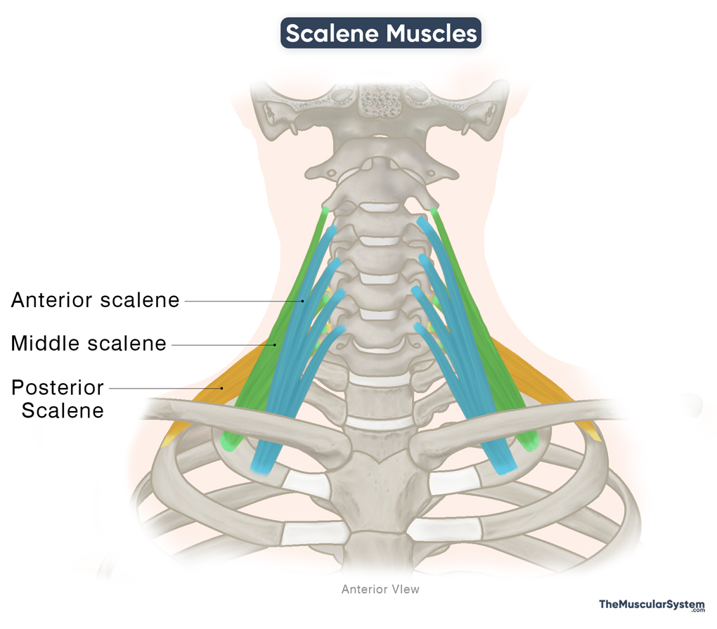
The scalene muscles are a group of three muscles located on the lateral sides of the neck, identified as the anterior, middle, and posterior scalenes. As part of the deep cervical muscles, the scalenes play a vital role in flexing and stabilizing the neck as well as elevating the upper ribs during respiration.
The name comes from the Ancient Greek word “skalēnós,” meaning “uneven,” referring to the shape of the muscle.
Anatomy
Location and Attachments
| Origin | Anterior: Transverse processes of the C3–C6 vertebrae Middle: Transverse processes of the C2-C7 vertebrae Posterior: Transverse processes of the C5–C7 vertebrae |
| Insertion | Anterior: Scalene tubercle on the upper surface of the 1st rib Middle: Superior surface of the 1st rib Posterior: External surface of the 2nd rib |
The three scalene muscles are named based on their points of origin in the cervical spine.
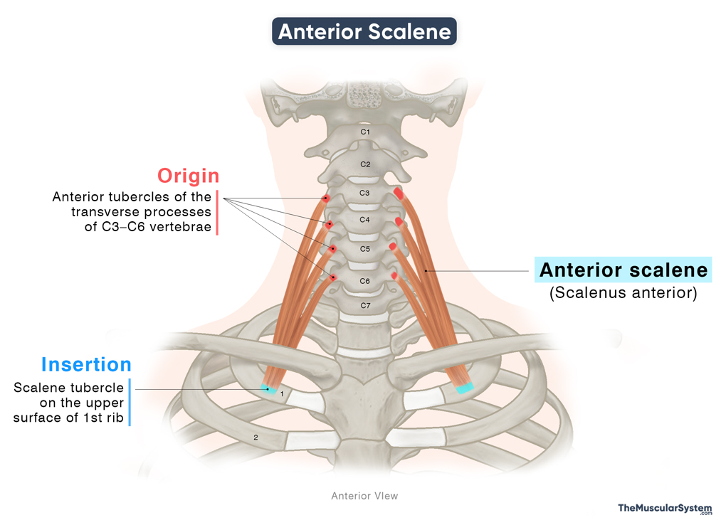
Anterior Scalene (Scalenus Anterior)
Origin: As the most anterior of the three scalene muscles, the anterior scalene arises from the anterior tubercles of the transverse processes of the third to sixth cervical vertebrae (C3–C6).
Insertion: The muscle descends vertically to reach the thoracic cage and inserts via a single tendon into the superior side of the first rib, mainly the scalene tubercle, a small bony ridge. This point of insertion lies just in front of the subclavian groove that allows passage to the subclavian artery.
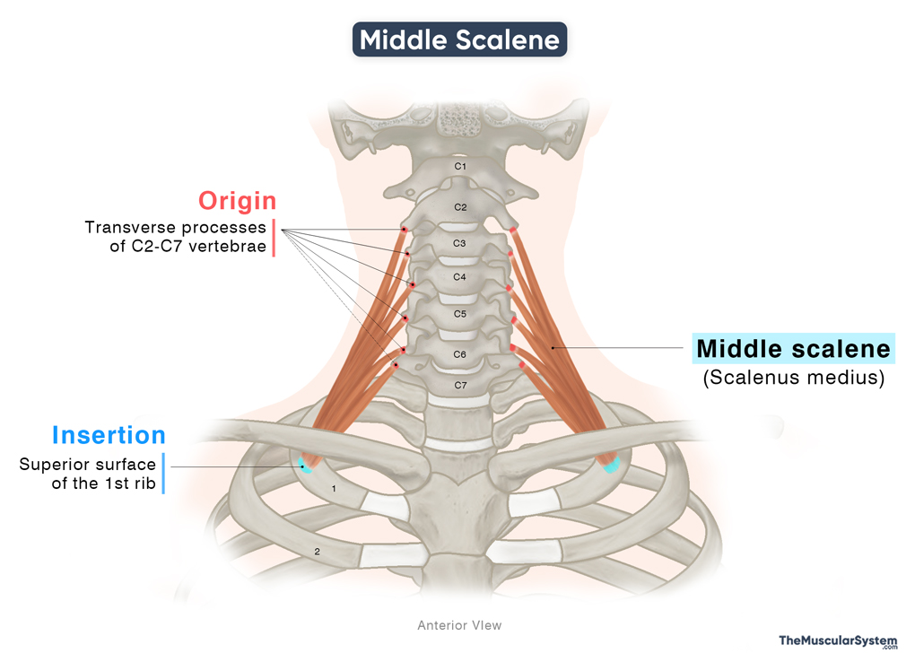
Middle Scalene (Scalenus Medius)
It is the longest of the three scalene muscles.
Origin: The muscle originates from the transverse processes of the axis (second cervical vertebra) to the seventh cervical vertebrae (C2-C7).
Insertion: From its origin, the muscle courses laterally toward the back, down along the vertebral column as it forms a single broad tendon. This tendon then inserts into the superior surface of the first rib, in front of the tubercle of the first rib, and behind the subclavian groove.
Posterior Scalene (Scalenus Posterior)
It is the smallest muscle in the group.
Origin: The most posterior of the three muscles, it arises via 2-3 tendinous bands from the posterior tubercles of the transverse processes of the fifth to seventh cervical vertebrae (C5–C7).
Insertion: Like the middle scalene, this muscle also descends posteriorly and laterally as the muscle belly narrows into a single tendon. It inserts into the external surface of the second rib. Its muscle fibers may sometimes blend with the middle scalene.

Relations With Surrounding Muscles and Structures
Muscular Relations
The anterior and middle scalene muscles lie deep to the sternocleidomastoid muscle and the prevertebral fascia. Their lower portions overlap with the subclavius and the inferior belly of the omohyoid.
The middle scalene is related laterally to the levator scapulae, which lies behind it. The posterior scalene muscle lies deep to the other scalenes, while the superior part of the serratus anterior lies lateral to it.
Neurovascular Relations
The anterior and middle scalene muscles form the front and back boundaries of the interscalene triangle, an opening located on the lower lateral side of the neck. The first rib forms the base of this triangle. Through this space passes the brachial plexus and the third part of the subclavian artery.
In front of the anterior scalene muscle lie the suprascapular, transverse cervical, and ascending cervical arteries, as well as the lateral aspect of the carotid sheath.
The dorsal scapular and long thoracic nerves pierce the lateral surface of the middle scalene muscle as they continue posteriorly to supply muscles of the back.
Variation
In some individuals, a few additional muscle slips arise from the anterior scalene, forming a fourth scalene muscle known as the scalenus minimus or scalenus pleuralis. This accessory muscle may insert into the suprapleural membrane and sometimes onto the inner surface of the first rib. Alternatively, it may originate directly from the transverse process of the seventh cervical vertebra (C7). The presence of this fourth scalene is highly variable, with studies reporting an incidence ranging from approximately 7% to 72% of the population.
Function
| Action | — Flexing and rotating the neck — Lifting the 1st-2nd ribs to assist during forced respiration |
Flexing the neck
- Bilateral contraction of the anterior scalene assists in forward neck flexion, bending the head forward.
- When all the scalenes contract unilaterally, they produce lateral flexion, bending the neck and head toward the same side as the contracting muscles.
- Unilateral contraction of the anterior scalene also helps rotate the head toward the opposite side.
Lifting the ribs to assist in respiration
The anterior, middle, and posterior scalene muscles contract along with the external intercostal muscles to help elevate the upper two ribs during inspiration. The anterior and middle scalenes elevate the first rib, while the posterior scalene elevates the second rib. Through this action, the scalenes function as accessory muscles of respiration, particularly during forced inspiration, as they help increase the volume of the thoracic cavity.
Antagonists
The semispinalis capitis, semispinalis cervicis, splenius capitis, and splenius cervicis act as antagonists to the scalene muscles during neck flexion, as they are the primary extensors of the cervical spine.
Innervation
| Nerve | Ventral rami of the third to eighth cervical spinal nerves (C3-C8) |
The three scalene muscles are innervated by the ventral rami of the cervical spinal nerves, with the specific nerve roots varying among the three:
- Anterior scalene: Fourth to sixth cervical spinal nerves (C4-C6)
- Middle scalene: Third to eighth cervical spinal nerves (C3-C8)
- Posterior scalene: Sixth to eighth cervical spinal nerves (C6-C8)
Blood Supply
| Artery | Ascending cervical artery |
All three scalene muscles receive their blood supply primarily from the ascending cervical artery, a branch of the inferior thyroid artery, which itself arises from the thyrocervical trunk of the subclavian artery.
Additional contributions may come from the transverse cervical artery, which also branches from the thyrocervical trunk.
References
- Anatomy, Head and Neck, Scalenus Muscle: NCBI.NLM.NIH.gov
- Scalene Muscles: Kenhub.com
- Scalenus Anterior Muscle: Elsevier.com
- Scalenus Medius Muscle: Elsevier.com
- Scalenus Posterior Muscle: Elsevier.com
- Scalene Muscles | Function, Innervation & Action: Study.com
- Scalenus Anterior Muscle: Radiopaedia.org
- Scalenus Medius Muscle: Radiopaedia.org
- Scalenus Posterior Muscle: Radiopaedia.org
Della Barnes, an MS Anatomy graduate, blends medical research with accessible writing, simplifying complex anatomy for a better understanding and appreciation of human anatomy.
- Latest Posts by Della Barnes, MS Anatomy
-
Tensor Tympani
- -
Stapedius
- -
Auricularis Posterior
- All Posts


