

Flexor Digitorum Longus
By
Della Barnes, an MS Anatomy graduate, blends medical research with accessible writing, simplifying complex anatomy for a better understanding and appreciation of human anatomy.
Last updated:
18/09/2025Della Barnes, MS Anatomy
UX/UI Designer at - AdobeDella Barnes, an MS Anatomy graduate, blends medical research with accessible writing, simplifying complex anatomy for a better understanding and appreciation of human anatomy.
What is the Flexor Digitorum Longus
The flexor digitorum longus (FDL), also called the flexor digitorum communis longus, is a long, thin muscle located on the tibial side of the posterior leg, running from the calf region to the plantar surface of the foot. It belongs to the deep posterior compartment of the lower leg, along with the flexor hallucis longus, tibialis posterior, and popliteus.
It is the primary muscle that acts to flex the second to fifth toes or digits, hence the name flexor “digitorum” longus.
Anatomy
Location and Attachments
| Origin | Posterior surface of the tibial body |
| Insertion | Distal phalangeal base of the 2nd to 5th toes (on the plantar surface) |
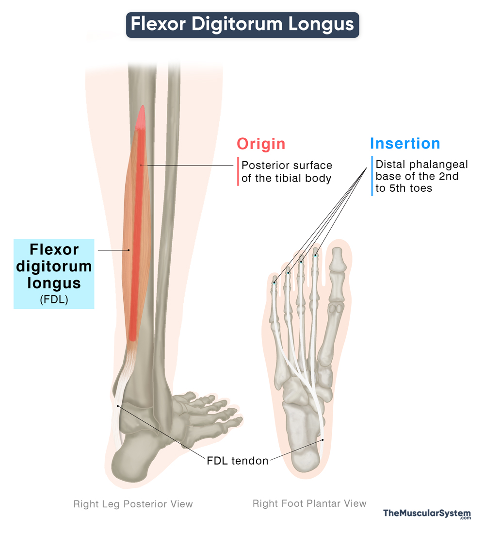
Origin
The flexor digitorum longus originates from the medial part of the posterior surface of the body of the tibia, just below the soleal line, which marks the origin of the soleus muscle. This point also lies medial to the origin of the tibialis posterior.
Insertion
From its origin, the muscle descends along the medial side of the lower leg toward the ankle joint. Just above the ankle, the fleshy belly tapers into a long, slender tendon. This tendon passes behind the medial malleolus of the tibia and beneath the flexor retinaculum, which secures it as it travels through the tarsal tunnel.
After leaving the tunnel, the tendon continues forward across the sole of the foot, angling slightly toward the lateral side. It then divides into four slender slips, each inserting into the base of the distal phalanx of the lateral four digits, which are the second through fifth toes.
Relations With Surrounding Muscles and Structures
Within the deep posterior compartment of the leg, the FDL lies at the back of the tibialis posterior and medial to the flexor hallucis longus. It lies deep to the muscles in the superficial posterior compartment, the gastrocnemius and soleus, and is separated from them by the deep fascia of the leg.
At the ankle, the FDL tendon crosses the tibialis posterior tendon just above the medial malleolus, a crossing referred to as the crural tendinous chiasm. Behind the medial malleolus, the order is Tibialis posterior, FDL, posterior tibial artery, tibial nerve, and FHL. In the sole, FDL crosses FHL at the knot of Henry, where FHL usually gives a slip to FDL.
Continuing into the sole, it passes the deltoid ligament superficially, and then crosses the tendon of the flexor hallucis longus (FHL) near the navicular bone. The two heads of the quadratus plantae attach to the FDL tendons, reinforcing their pull and assisting in toe flexion. The lumbrical muscles also arise from these tendons, contributing to toe stabilization and fine motor control. Distally, the four tendons of the flexor digitorum longus pass deep to the corresponding tendons of the flexor digitorum brevis before inserting into the distal phalanges.
Function
| Action | Flexing the 2nd to 4th toes at all their joints, and helping with plantar flexion |
It is the primary muscle responsible for flexing the second through fifth toes at both the metatarsophalangeal and interphalangeal joints. It also assists in plantar flexion of the foot at the ankle joint. By curling the lesser toes, the muscle enables the foot to maintain a firm grip on the ground, helping with maintaining balance on irregular or uneven surfaces.
During walking, the muscle’s activity is closely coordinated with the gait cycle. As the foot is lifted, the flexor digitorum longus bends the toes in sequence, beginning at the distal joints and progressing proximally. Once the foot makes contact with the ground, the muscle works in synergy with the lumbricals and interossei to keep the toes firmly on the surface, stabilizing the foot and supporting balance.
Because its tendon passes behind the medial malleolus, the flexor digitorum longus also contributes to inversion of the foot at the subtalar joint (between the talus and calcaneus). This is a secondary action, carried out alongside the tibialis anterior and tibialis posterior, which are the principal muscles for Inversion of the foot.
Antagonists
The primary antagonists to the flexor digitorum longus muscle are the extensor digitorum longus and extensor digitorum brevis muscles, which extend the second through fifth toes in opposition to its flexion.
Innervation
| Nerve | Tibial nerve (L5-S2) |
The muscle receives innervation from the tibial nerve, arising from the fifth lumbar and first to second sacral nerve roots (L5-S2). The tibial nerve itself is a branch of the sciatic nerve.
Blood Supply
| Artery | Posterior tibial artery |
Blood supply to the muscle comes from the posterior tibial artery, one of the two terminal branches of the popliteal artery.
References
- Flexor Digitorum Longus: Elsevier.com
- Flexor Digitorum Longus Muscle: Radiopaedia.org
- Flexor Digitorum Longus Muscle: Kenhub.com
- Flexor Digitorum Longus Muscle – Attachments, Actions & Innervation: GetBodySmart.com
- Flexor Digitorum Longus: TeachMeAnatomy.info
Della Barnes, an MS Anatomy graduate, blends medical research with accessible writing, simplifying complex anatomy for a better understanding and appreciation of human anatomy.
- Latest Posts by Della Barnes, MS Anatomy
-
Tensor Tympani
- -
Stapedius
- -
Auricularis Posterior
- All Posts


