

Vastus Lateralis
By
Della Barnes, an MS Anatomy graduate, blends medical research with accessible writing, simplifying complex anatomy for a better understanding and appreciation of human anatomy.
Last updated:
22/08/2025Della Barnes, MS Anatomy
UX/UI Designer at - AdobeDella Barnes, an MS Anatomy graduate, blends medical research with accessible writing, simplifying complex anatomy for a better understanding and appreciation of human anatomy.
What is the Vastus Lateralis
The vastus lateralis, sometimes called the vastus externus, is a large, unipennate muscle located on the lateral side of the thigh. It is one of the four quadriceps muscles, along with the rectus femoris, vastus medialis, and vastus intermedius, and is the largest among them. Together with the other quadriceps muscles and the sartorius, it forms the anterior compartment of the thigh.
Its muscle fibers are arranged diagonally on one side of a central tendon, characteristic of a unipennate muscle. It extends from the proximal femur to the top of the knee joint capsule and plays a vital role in knee extension.
Anatomy
Location and Attachments
| Origin | The intertrochanteric line, greater trochanter, gluteal tuberosity, and linea aspera of the femur |
| Insertion | — The base of the patella (kneecap) via the quadriceps femoris tendon — The tibial tuberosity via the patellar ligament |
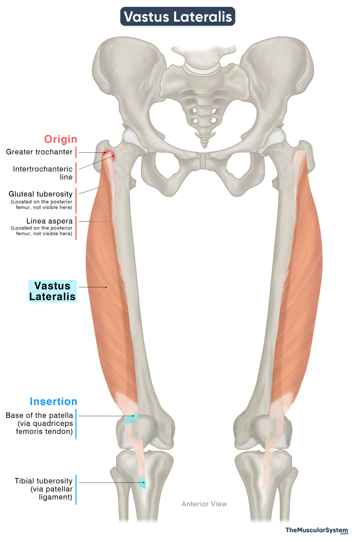
Origin
The muscle originates via a broad attachment to the proximal femur and the surrounding fascia. The primary points of attachment include:
- The upper border of the intertrochanteric line
- The front aspect of the greater trochanter
- The gluteal tuberosity of the femur
- The outer lip of the linea aspera of the femur
- The adjacent part of the lateral intermuscular septum
Insertion
From their point of origin, the tendons thicken to form a large and fleshy muscle belly. It descends along the lateral sides of the anterior thigh and converges into a flat, broad aponeurotic tendon. This tendon joins with those of the rectus femoris, vastus medialis, and vastus intermedius to form the quadriceps tendon. The quadriceps tendon inserts into the base (superior surface) of the patella.
From the apex (inferior surface) of the patella, some part of the quadriceps tendon continues as the patellar ligament or patellar tendon and attaches to the tibial tuberosity on the front of the tibia. Through this connection, the vastus lateralis, along with the other quadriceps muscles, exerts force on the tibia to extend the knee.
Relations With Surrounding Muscles and Structures
The vastus lateralis is the most lateral of the four quadriceps muscles. Medially, it is bordered by the rectus femoris and the vastus intermedius. The lateral femoral circumflex artery and branches of the femoral nerve pass between the vastus lateralis and vastus intermedius.
Anteriorly, the muscle lies deep to the skin, superficial fascia, and the fascia lata, the deep fascia that envelops all thigh muscles.
Lateral to the muscle are the muscles tensor fasciae latae, gluteus maximus, and the iliotibial tract, all of which lie superficial to it.

Posteriorly, the vastus lateralis is separated from the long and short heads of the biceps femoris, a muscle of the posterior thigh, by the lateral intermuscular septum, a fascial partition that demarcates the anterior and posterior compartments of the thigh.
Function
| Action | Extending the knee |
The Vastus Lateralis muscle works on the knee joint with two main functions:
Extension: Being the largest of the quadriceps muscles, it is a major extensor of the lower leg at the knee joint, which makes the muscle instrumental for all kinds of leg movements, like walking, climbing, running, sitting down, and standing up.
Stabilization: It contributes to stabilizing the knee joint by helping the patella (kneecap) to stay in place within the femoral groove. The vastus lateralis maintains a lateral pull on the patella, which balances the medial pull mainly from the vastus medialis. This action reinforces the stability of the knee joint during movement.
Antagonists
The hamstring muscles, biceps femoris, semitendinosus, and semimembranosus, act as antagonists to the vastus lateralis by producing knee flexion, which opposes its role in knee extension.
Innervation
| Nerve | Femoral nerve (L2-L4) |
The muscle gets its innervation from the largest branch of the lumbar plexus, the femoral nerve, which rises from the L2 to L4 nerve roots.
Blood Supply
| Artery | Lateral circumflex femoral artery |
Blood supply to the muscle comes from the transverse and descending branches of the lateral circumflex femoral artery, a branch of the deep femoral artery. Additional blood supply may be provided by the perforating arteries of the deep femoral artery.
References
- Anatomy, Bony Pelvis and Lower Limb: Vastus Lateralis Muscle: NCBI.NLM.NIH.gov
- Vastus Lateralis Muscle: Elsevier.com
- Vastus Lateralis: TeachMeAnatomy.info
- Vastus Lateralis Muscle: GetBodySmart.com
- Quadriceps Femoris Muscle: Kenhub.com
- Vastus lateralis: HealthLine.com
Della Barnes, an MS Anatomy graduate, blends medical research with accessible writing, simplifying complex anatomy for a better understanding and appreciation of human anatomy.
- Latest Posts by Della Barnes, MS Anatomy
-
Tensor Tympani
- -
Stapedius
- -
Auricularis Posterior
- All Posts


