

Occipitalis
By
Della Barnes, an MS Anatomy graduate, blends medical research with accessible writing, simplifying complex anatomy for a better understanding and appreciation of human anatomy.
Last updated:
31/01/2026Della Barnes, MS Anatomy
UX/UI Designer at - AdobeDella Barnes, an MS Anatomy graduate, blends medical research with accessible writing, simplifying complex anatomy for a better understanding and appreciation of human anatomy.
The occipitalis is a quadrilateral muscle located at the back of the skull. This muscle, together with the frontalis, forms the occipitofrontalis, an important muscular structure that covers the skull and helps with facial expressions.
Anatomy
Location and Attachments
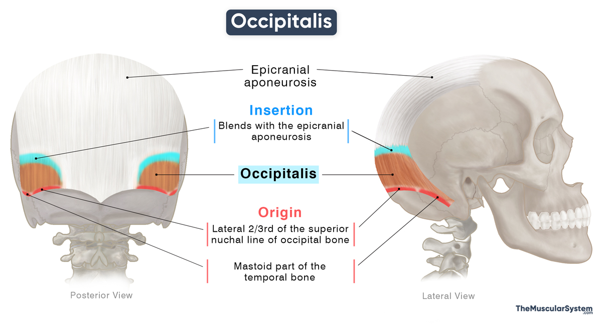
| Origin | — Lateral 2/3 of the superior nuchal line of the occipital bone — Mastoid part of the temporal bone |
| Insertion | The epicranial aponeurosis |
Origin
The muscle has a broad tendinous origin along the occipital bone’s superior nuchal line, typically covering its lateral two-thirds. The point of origin continues laterally further to include the adjacent portion of the mastoid process of the temporal bone.
Insertion
The muscle fibers course superiorly as they form the short and thin muscle belly. The muscle then inserts into the epicranial aponeurosis.
Relations With Surrounding Muscles and Structures
Since there are not many muscles in the scalp, the occipitalis lies directly over the occipital bone, forming the superficial muscular layer at the back of the skull. At its bony origin along the superior nuchal line and mastoid region, the muscle overlies and sits just superior to the cranial attachments of deeper posterior neck muscles, primarily the semispinalis capitis, splenius capitis, and the upper fibers of trapezius.
The epicranial aponeurosis, where the muscle inserts, is the fibrous aponeurotic layer that covers the top of the skull, connecting the occipitalis with the frontalis, enabling coordinated anterior-posterior movement of the scalp
The occipital artery and the greater occipital nerve cross the superficial surface of the muscle.
Function
| Action | Retracting the forehead and helping with facial expressions |
The occipitalis retracts or pulls the scalp back towards the occipital bone, tightening and stabilizing the epicranial aponeurosis. This action counterbalances the forward pull of the frontalis, helping it contract more effectively to pull up the forehead. Together, the two muscles coordinate to produce expressions such as raised eyebrows and horizontal forehead wrinkles.
In some people, its contraction may activate the nearby auricular muscles, allowing them to wiggle their ears slightly.
Antagonists
It does not have a direct antagonist because returning the scalp to its resting position after being pulled backward does not require an opposing muscle.
Innervation
| Nerve | Occipital branch of the posterior auricular nerve |
The muscle is innervated by the occipital branch of the posterior auricular nerve, which rises from the facial nerve (CN VII).
Blood Supply
| Artery | Occipital artery and posterior auricular artery |
Blood supply to this muscle comes from the occipital artery, while the posterior auricular artery provides additional vasculature. Both are branches of the external carotid artery.
References
- Occipitalis Muscle | Function, Origin & Insertion: Study.com
- Occipitalis Muscle: Elsevier.com
- Occipitalis Muscle: Radiopaedia.org
- Occipitalis Muscle: Kenhub.com
- Occipitalis Muscle: IMAIOS.com
Della Barnes, an MS Anatomy graduate, blends medical research with accessible writing, simplifying complex anatomy for a better understanding and appreciation of human anatomy.
- Latest Posts by Della Barnes, MS Anatomy
-
Tensor Tympani
- -
Stapedius
- -
Auricularis Posterior
- All Posts


