

Frontalis
By
Della Barnes, an MS Anatomy graduate, blends medical research with accessible writing, simplifying complex anatomy for a better understanding and appreciation of human anatomy.
Last updated:
31/01/2026Della Barnes, MS Anatomy
UX/UI Designer at - AdobeDella Barnes, an MS Anatomy graduate, blends medical research with accessible writing, simplifying complex anatomy for a better understanding and appreciation of human anatomy.
The frontalis is a thin, broad muscle that overlies the frontal bone and forms the anterior component of the occipitofrontalis. It plays a key role in elevating the eyebrows and wrinkling the forehead, making it essential for many facial expressions.
The muscle is a common target for Botox injections, as its repetitive contraction is responsible for the formation of horizontal forehead lines.
Anatomy
Interestingly, the muscle has no direct bony attachments, which gives it considerable mobility as it lies superficially over the frontal bone, forming most of the forehead.
Location and Attachments
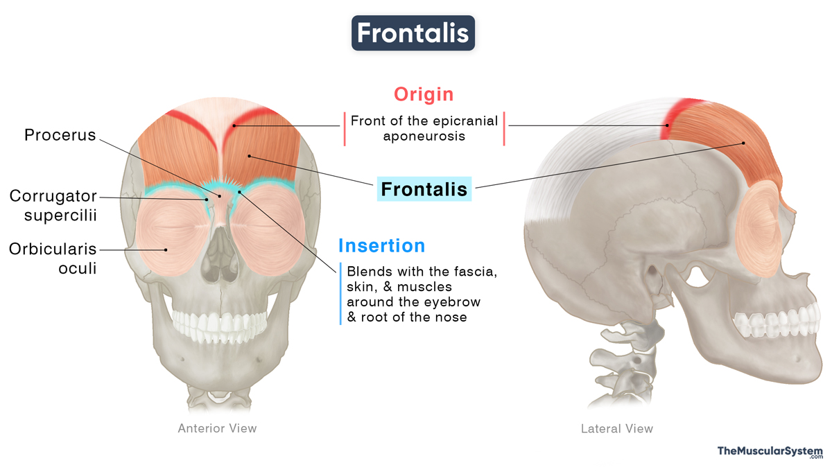
| Origin | Front of the epicranial aponeurosis |
| Insertion | The fascia and skin around the eyebrow, and the root of the nose |
Origin
The muscle originates via tendinous fibers from the front of the epicranial aponeurosis. It is the fibrous aponeurosis that covers the top of the skull, mainly overlying the parietal bones.
Insertion
The muscle fibers form a thin, broad belly that descends over the skull, down the forehead, and attaches to the superficial fascia and skin around the eyebrows. Some of the fibers also extend further down to insert into the root of the nose.
Relations With Surrounding Muscles and Structures
Lying immediately beneath the superficial fascia of the scalp, the frontalis muscle is broader than the occipitalis and composed of longer, paler fibers. The two muscles are connected across the cranium through the epicranial aponeurosis. The frontalis arises near the level of the coronal suture, a major cranial suture separating the frontal and parietal bones.
As it approaches its distal attachment near the eyebrows, the frontalis blends seamlessly with adjacent facial muscles. Laterally, it blends with the eyelid muscles, the corrugator supercilii and orbicularis oculi. On the medial side, its fibers merge with the procerus muscle of the nose.
Function
| Action | Raising the eyebrows and helping wrinkle the forehead to express surprise or worry |
When the muscle contracts, it pulls on the skin of the forehead, helping with a range of facial expressions. Key actions include:
- Lifting Eyebrows: Conveys emotions like surprise and shock.
- Forehead Wrinkling: Creates horizontal lines to express worry or concern
Since the muscle has two separate heads, it allows for these movements to occur on one side of the face, such as raising one eyebrow in surprise.
Antagonists
The orbicularis oculi, being the primary muscle that closes the eyelids, is antagonistic to the frontalis muscle. The corrugator supercilii and procerus, which serve as the key frowning muscles, also oppose the frontalis by drawing the eyebrows downward and medially to produce vertical forehead lines.
Innervation
| Nerve | Temporal branch of facial nerve (CN VII) |
The muscle is innervated by the temporal branch of the facial nerve (CN VII). It is the most superior of the five terminal motor nerve branches of the facial nerve.
Blood Supply
| Artery | Supraorbital and supratrochlear arteries |
Blood supply to the muscle is provided primarily by the supraorbital artery, one of the orbital branches of the ophthalmic artery, and the supratrochlear artery, one of its terminal branches. The ophthalmic artery is a branch of the internal carotid artery.
The frontal branch of the superficial temporal artery, arising from the external carotid artery, may also provide additional vascularization.
References
- Frontalis Muscle | Location, Function & Action: Study.com
- Frontalis Muscle: Kenhub.com
- Anatomy, Head and Neck; Frontalis Muscle: NCBI.NLM.NIH.gov
- Frontalis Muscle: Elsevier.com
- Frontalis Muscle – Attachments, Actions & Innervation: GetBodySmart.com
Della Barnes, an MS Anatomy graduate, blends medical research with accessible writing, simplifying complex anatomy for a better understanding and appreciation of human anatomy.
- Latest Posts by Della Barnes, MS Anatomy
-
Tensor Tympani
- -
Stapedius
- -
Auricularis Posterior
- All Posts


