

Orbicularis Oculi
By
Della Barnes, an MS Anatomy graduate, blends medical research with accessible writing, simplifying complex anatomy for a better understanding and appreciation of human anatomy.
Last updated:
10/02/2026Della Barnes, MS Anatomy
UX/UI Designer at - AdobeDella Barnes, an MS Anatomy graduate, blends medical research with accessible writing, simplifying complex anatomy for a better understanding and appreciation of human anatomy.
The orbicularis oculi is a paired circular muscle that forms a muscular loop or ring around the eye socket or orbit. This is the muscle primarily responsible for closing the eyelids. It is one of the vital muscles for facial expression, along with its surrounding muscles, such as the corrugator supercilii, frontalis, and levator palpebrae superioris.
Anatomy
The sphincter-like muscle lies just beneath the skin around the eyes. It surrounds the orbit, taking a continuous elliptical form, covering the entire circumference around each eye. It has several divisions and subdivisions based on their attachments.
Location and Attachments
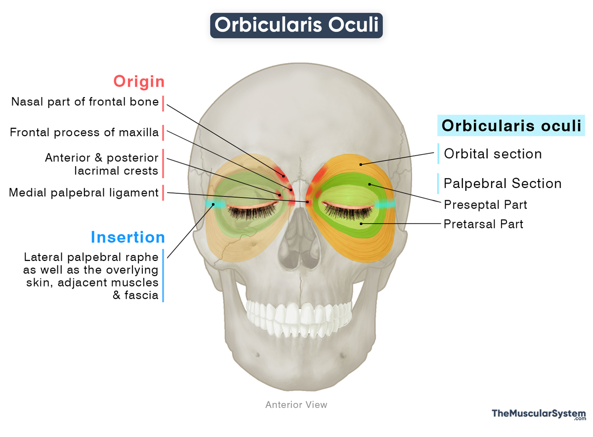
| Origin | — The nasal part of the frontal bone — The frontal process of the maxilla — The medial palpebral ligament — The anterior and posterior lacrimal crests |
| Insertion | The lateral palpebral raphe, the overlying skin, and the adjacent muscles and fascia |
The Orbital Section
Origin
It is the largest and thickest part of the muscle, characterized by darker, reddish fibers. It arises from the medial side of the bony orbit, specifically from the nasal part of the frontal bone and the frontal process of the maxilla.
A small portion may also arise from the front of the medial palpebral ligament, the ligament at the inner corner of the eye that anchors the skin (medial canthus) to the bony orbit.
Insertion
From their origin, the fibers curve laterally in a continuous loop around the orbital margin, extending above and below the eye. Along their course, they blend with adjacent muscles, connective tissues, and the overlying skin. The superior and inferior fibers ultimately converge at the lateral palpebral raphe, a fibrous band created by the interlacing lateral fibers of the orbicularis oculi.
The Palpebral Section
Origin
Although a few orbital fibers can arise from the medial palpebral ligament, this ligament serves primarily as the origin of the palpebral portion of the orbicularis oculi. The palpebral fibers are thinner and paler than those of the orbital section, and consist of two subdivisions, each with two distinct heads.
Preseptal Part
- The superficial head arises from the front surface of the medial palpebral ligament.
- The deep head originates from the posterior lacrimal crest on the lacrimal bone and the fibrous connective tissue surrounding the lacrimal sac.
Pretarsal Part
- The superficial head originates from the anterior lacrimal crest on the maxilla.
- The deep head, commonly known as the Horner’s muscle, or the lacrimal part, arises from the posterior lacrimal crest (lacrimal bone) and the medial palpebral ligament.
Insertion
Preseptal Part: The fibers loop around the orbit and insert into the lateral orbital or Whitnall’s tubercle on the orbital surface of the zygomatic bone.
Pretarsal Part: Like the preseptal part, the fibers of this portion firmly encircle the upper and lower eyelids, inserting into the lateral palpebral raphe.
Some sources describe a separate lacrimal section of the muscle; however, it is considered part of the palpebral section here.
The Ciliary Section (Muscle of Riolan)
The marginal fibers of the palpebral part of the muscle run along the very edge of both upper and lower eyelids, forming the grayish line easily visible along the eyelids. This part is often referred to as Riolan’s muscle or the ciliary bundle. It is an important anatomical and surgical landmark.
Relations With Surrounding Muscles and Structures
Muscular Relations
It blends with the surrounding muscles, extending to the eyebrows, and adjacent temporal and infraorbital regions. Superiorly, some of its fibers merge with various forehead and eyebrow muscles, including the occipitofrontalis, corrugator supercilii, and depressor supercilii.
Medially and inferiorly, the orbital fibers may intermingle with muscles of the upper lip and nasolabial area, including levator labii superioris and zygomaticus minor. When present, the cheekbone muscle, malaris, also merges with these lower fibers.
Neurovascular Relations
The palpebral branches of the infraorbital nerve pierce the deep surface of the muscle. The supraorbital vein and the zygomaticofacial nerve also pass directly through the orbital portion of the muscle as they course toward the forehead and the upper cheek.
The fibers of levator palpebrae superioris pierce the upper part of orbicularis oculi on their way to the skin of the upper eyelid.
Function
The orbicularis oculi contains about 88% fast-twitch (type II) fibers and 12% slow-twitch (type I) fibers. The high percentage of fast-twitch fibers allows the muscle to perform rapid, reflexive actions like blinking and quick eye closure, while the smaller slow-twitch portion supports gentle, sustained eyelid closure, such as keeping the eyes closed without fatigue during sleep.
| Action | Controlling the closing of the eyelids and helping with better tear drainage |
1. Forceful Closing of the Eyes (Orbital Section)
Being the thicker, peripheral portion of the muscle that blends with the surrounding skin and facial muscles, it is responsible for the forceful closure of the eyes.
How it Works: When this part contracts, it tightens like a strong sphincter, pulling the nearby skin and muscles toward the orbital rim and clamping the eyelids firmly together. This action is used when squinting against bright light or when closing the eyes tightly to resist a sudden blow.
2. Gentle Closing of the Eyes (Palpebral Section)
This portion of the muscle handles the regular, gentle closing of the eyes, such as during blinking and sleeping.
How it Works: Its fibers run horizontally across the eyelids from the medial corner of the eye (medial palpebral ligament) to the lateral corner (lateral palpebral raphe). When these fibers contract, they shorten the eyelids in the medial-to-lateral direction, drawing the upper and lower eyelids toward each other in a smooth, gliding motion.
These movements are usually involuntary, but they can occur voluntarily as a mild protective reflex against irritants like wind or dust. As the upper and lower eyelids meet, it spreads the tear from the lacrimal gland over the cornea, helping clear debris and keep the eye moist. This is why closing your eyes against a gust of wind often brings out tears, to wash away any dust that entered.
It is the primary muscle responsible for closing the eyelids, and damage to this muscle can result in an inability to close the eye properly, often requiring protective or corrective surgical procedures.
3. Tear Drainage (Palpebral Section)
The lacrimal part, or Horner’s muscle, is vital in the proper drainage of tears.
How it Works: The deep fibers of the muscle that attach to the lacrimal bone and surround the lacrimal sac contract to pull the sac laterally, briefly enlarging it like a small suction pump. This creates negative pressure inside the sac, helping tears enter it from the canaliculi.
When the muscle relaxes, the sac recoils and pushes the tears down into the nasolacrimal duct, improving overall tear drainage with each blink.
Antagonists
The levator palpebrae superioris, the extraocular muscle responsible for lifting the upper eyelid to open the eye, acts as the antagonist to the orbicularis oculi, which closes it.
Innervation
| Nerve | temporal and zygomatic branches of the facial nerve |
The facial nerve (CN VII) innervates this muscle via its temporal and zygomatic branches.
Blood Supply
| Artery | Aygomatico-orbital and angular arteries (external carotid artery), and the ophthalmic artery (internal carotid artery) |
Blood supply to the muscle comes from the superficial temporal artery, via the zygomatico-orbital branch of the middle temporal artery. The angular artery, the terminal branch of the facial artery, also provides vasculature to this muscle. Both these sources come from the external carotid artery.
Another vital source of blood supply is the ophthalmic artery, a branch of the internal carotid artery.
References
- Anatomy, Head and Neck: Orbicularis Oculi Muscle: NCBI.NLM.NIH.gov
- Orbicularis Oculi: TeachMeAnatomy.info
- Orbicularis Oculi Muscle | Function, Origin & Insertion: Study.com
- Orbicularis Oculi: Kenhub.com
- Orbicularis Oculi Muscle (Left): Elsevier.com
Della Barnes, an MS Anatomy graduate, blends medical research with accessible writing, simplifying complex anatomy for a better understanding and appreciation of human anatomy.
- Latest Posts by Della Barnes, MS Anatomy
-
Tensor Tympani
- -
Stapedius
- -
Auricularis Posterior
- All Posts


