

Pharyngeal Muscles
By
Della Barnes, an MS Anatomy graduate, blends medical research with accessible writing, simplifying complex anatomy for a better understanding and appreciation of human anatomy.
Last updated:
05/03/2026Della Barnes, MS Anatomy
UX/UI Designer at - AdobeDella Barnes, an MS Anatomy graduate, blends medical research with accessible writing, simplifying complex anatomy for a better understanding and appreciation of human anatomy.
The pharynx is the part of the upper gastrointestinal tract between the oral (and nasal) cavities and the esophagus. It is divided into three parts: the oropharynx, nasopharynx, and laryngopharynx. A group of six skeletal muscles, referred to as the pharyngeal muscles, forms the walls of the pharynx.
Apart from contributing to the pharyngeal wall, these muscles are instrumental in the functioning of the pharynx, including swallowing and vocalization.
Names and Anatomy of the Muscles of the Pharynx
There are six pharyngeal muscles, divided into two groups based on their fibers’ orientation: the superficial circular layer and the deep longitudinal layer. The coordinated contraction and relaxation of these muscles play a vital role in swallowing as they constrict, widen, and shorten the pharynx to lead the chewed food, or bolus, from the oral cavity to the esophagus, via the larynx.
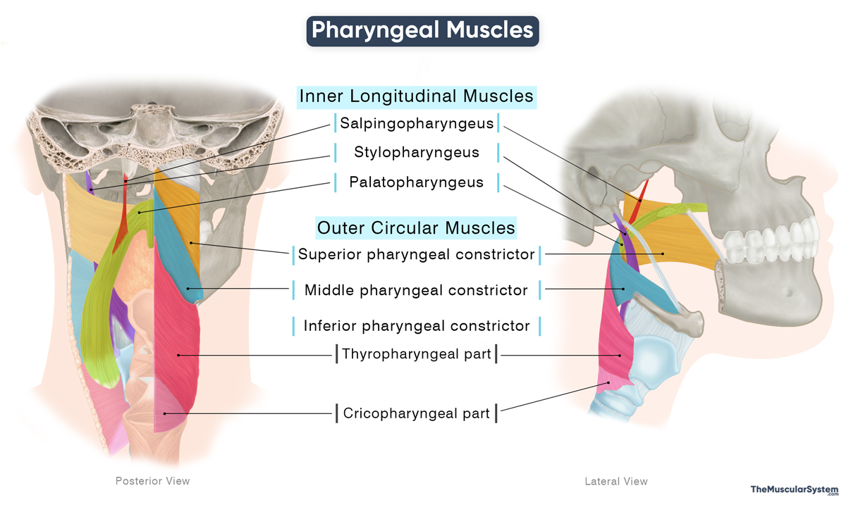
Here is a list of the pharyngeal muscles with their anatomy and actions:
| Name | Origin | Insertion | Action | Innervation | Blood Supply |
|---|---|---|---|---|---|
| Outer Circular Layer Muscles with fibers encircling the pharynx that contract sequentially to constrict the pharynx and propel food. | |||||
| Superior Pharyngeal Constrictor | Pterygoid hamulus, pterygomandibular raphe, back of the mylohyoid line, and sides of the tongue | Median pharyngeal raphe | Constricting the upper portion of the pharynx to help propel the bolus down the pharynx during swallowing | Pharyngeal plexus of the vagus nerve (CN X) | Pharyngeal trunk of the ascending pharyngeal artery and tonsillar artery |
| Middle Pharyngeal Constrictor | Greater and lesser horns of the hyoid bone, and the stylohyoid ligament | Median pharyngeal raphe | Constricting the middle portion of the pharynx to help propel the bolus further down | Pharyngeal plexus of the vagus nerve (CN X) | Pharyngeal trunk of the ascending pharyngeal artery and tonsillar artery |
| Inferior Pharyngeal Constrictor | Oblique line of the thyroid cartilage and the cricoid cartilage | Median pharyngeal raphe | Constricting the lowest portion of the pharynx to propel the bolus into the esophagus | Pharyngeal plexus of the vagus nerve (CN X) | Ascending pharyngeal and inferior thyroid arteries |
| Inner longitudinal Layer Muscles that have fibers that run lengthwise, shortening and elevating the pharynx to guide food into the esophagus. | |||||
| Stylopharyngeus | Medial base of the temporal styloid process | Posterior margin of the thyroid cartilage, lateral glossoepiglottic fold, superior and middle pharyngeal constrictors | — Elevating the pharynx and larynx to enlarge the pharyngeal opening to receive the bolus — Helping with vocalization | Glossopharyngeal nerve (CN IX) | Pharyngeal trunk of the ascending pharyngeal artery |
| Salpingopharyngeus | The inferior cartilaginous portion of the Eustachian tube | Palatopharyngeus muscle | — Elevating the pharynx and larynx so the bolus can reach the esophagus — Helping with vocalization — Helping open the Eustachian tube to equalize air pressure | Pharyngeal plexus of the vagus nerve (CN X) | Ascending palatine artery, greater palatine artery, and pharyngeal trunk of the ascending pharyngeal artery |
| Palatopharyngeus | Back of the hard palate, and palatine aponeurosis of the soft palate | Posterior margin of the thyroid cartilage, and the sides of the pharynx | — Pulling the pharynx superiorly, medially, and anteriorlyto help with swallowing — Tensing the soft palate | Pharyngeal plexus of the vagus nerve (CN X) | Ascending palatine artery, greater palatine artery, and pharyngeal trunk of the ascending pharyngeal artery |
Stylopharyngeus is the only pharyngeal muscle that does not receive motor innervation from the vagus nerve.
The palatopharyngeus is considered both a pharyngeal muscle and a muscle of the soft palate, as it originates from the palatine aponeurosis and directly elevates and tenses the soft palate during swallowing. It contributes to the palatopharyngeal arch, a mucosal fold extending from the soft palate to the pharyngeal wall.
References
- Anatomy, Head and Neck: Pharyngeal Muscles: NCBI.NLM.NIH.gov
- Muscles and Walls of the Pharynx: Kenhub.com
- The Pharynx: TeachMeAnatomy.info
- Pharyngeal Muscles: Radiopaedia.org
Della Barnes, an MS Anatomy graduate, blends medical research with accessible writing, simplifying complex anatomy for a better understanding and appreciation of human anatomy.
- Latest Posts by Della Barnes, MS Anatomy
-
Tensor Tympani
- -
Stapedius
- -
Auricularis Posterior
- All Posts


