

Vastus Intermedius
By
Della Barnes, an MS Anatomy graduate, blends medical research with accessible writing, simplifying complex anatomy for a better understanding and appreciation of human anatomy.
Last updated:
19/08/2025Della Barnes, MS Anatomy
UX/UI Designer at - AdobeDella Barnes, an MS Anatomy graduate, blends medical research with accessible writing, simplifying complex anatomy for a better understanding and appreciation of human anatomy.
What is the Vastus Intermedius
The vastus intermedius is a broad, long, bipennate muscle in the anterior compartment of the thigh. It lies at the front of the thigh, behind the rectus femoris muscle, and is one of the four muscles constituting the quadriceps femoris complex, along with rectus femoris, vastus lateralis, and vastus medius.
Anatomy
Location and Attachments
| Origin | Anterior surface of the upper two-thirds of the femoral shaft |
| Insertion | — The base of the patella (kneecap) via the quadriceps femoris tendon — The tibial tuberosity via the patellar ligament |
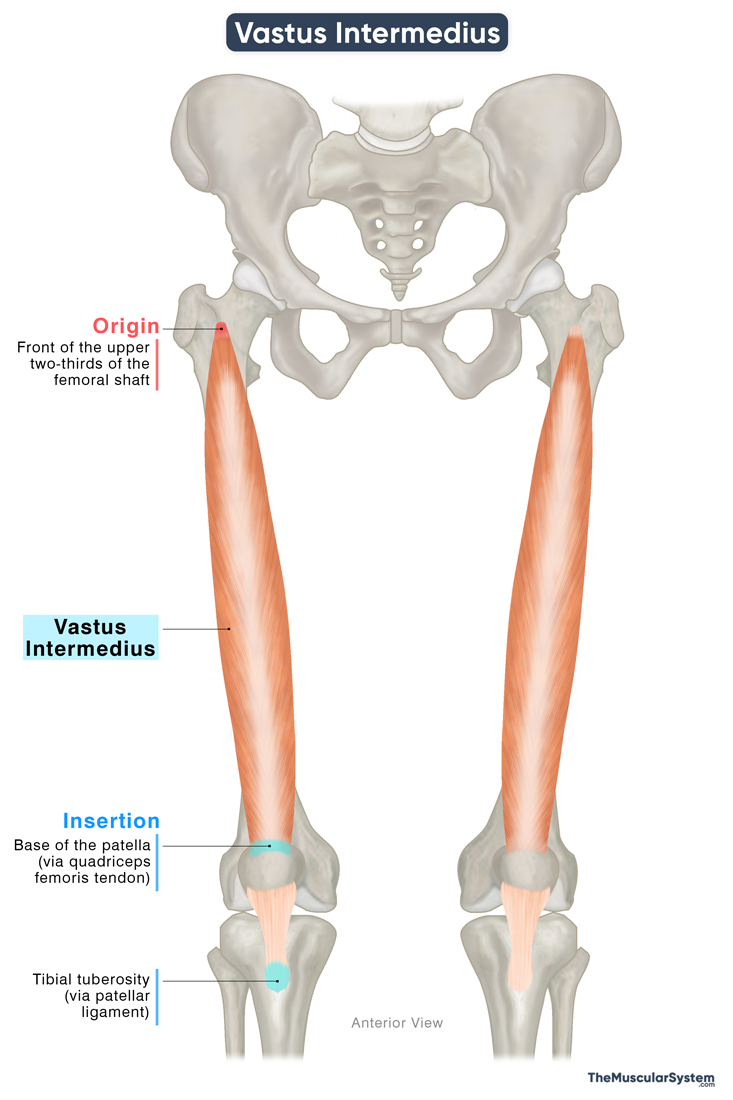
Origin
The muscle originates from the proximal femur, just inferior to the femoral neck, with fibers arising from the superior two-thirds of the anterior femoral shaft. Some muscle fibers may also originate from the lateral side of the femoral shaft and the lateral intermuscular septum.
Insertion
From its origin, the muscle descends almost vertically along the length of the femur. The muscle belly has a bipennate structure, meaning its fibers are arranged obliquely along a central tendon, resembling the shape of a feather.
As it reaches the distal end of the femur, the muscle belly becomes aponeurotic and merges with the quadriceps femoris tendon — the common tendon of the quadriceps group. This tendon then inserts into the superior surface or the base of the patella.
Along the patella, some part of the quadriceps tendon becomes continuous with the patellar ligament and inserts into the tibial tuberosity. This attachment establishes the connection between the quadriceps muscles and the tibia through which the muscles act on the lower leg.
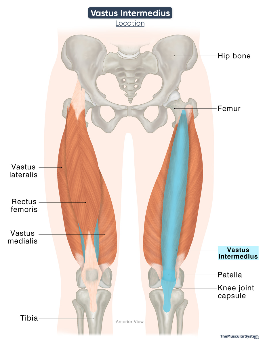
Relations With Surrounding Muscles and Structures
The vastus intermedius gets its name from its location between the vastus lateralis and vastus medialis. It is the deepest muscle in the anterior compartment of the thigh, with the femur lying directly behind it. The rectus femoris, the most superficial thigh muscle, lies in front of the vastus intermedius, covering it completely.
Function
| Action | Extending the knee |
As part of the quadriceps group, the primary function of the muscle is to extend the lower leg at the knee joint. It means the muscle is vital for actions like walking, climbing, and standing up from a seated position.
Antagonists
The hamstring muscles, biceps femoris, semitendinosus, and semimembranosus, flex the knee joint, making them antagonistic to the vastus intermedius as a quadriceps muscle.
Innervation
| Nerve | Femoral nerve (L2-L4) |
Like the other quadriceps muscles, the vastus intermedius is innervated by muscular branches of the femoral nerve, which originates from the L2 to L4 spinal nerve roots. The femoral nerve is the largest branch of the lumbar plexus.
Blood Supply
| Artery | Lateral circumflex femoral artery |
The primary blood supply to the muscle comes from the lateral circumflex femoral artery, which is a branch of the deep femoral artery.
References
- Vastus Intermedius: TeachMeAnatomy.info
- Vastus Intermedius Muscle: Elsevier.com
- Vastus Intermedius Muscle: ScienceDirect.com
- Vastus Intermedius Muscle: Radiopaedia.org
- Vastus Intermedius: Rad.UW.edu
- Quadriceps Femoris Muscle: Kenhub.com
Della Barnes, an MS Anatomy graduate, blends medical research with accessible writing, simplifying complex anatomy for a better understanding and appreciation of human anatomy.
- Latest Posts by Della Barnes, MS Anatomy
-
Tensor Tympani
- -
Stapedius
- -
Auricularis Posterior
- All Posts


