

Splenius Cervicis
By
Della Barnes, an MS Anatomy graduate, blends medical research with accessible writing, simplifying complex anatomy for a better understanding and appreciation of human anatomy.
Last updated:
07/10/2024Della Barnes, MS Anatomy
UX/UI Designer at - AdobeDella Barnes, an MS Anatomy graduate, blends medical research with accessible writing, simplifying complex anatomy for a better understanding and appreciation of human anatomy.
What is the Splenius Cervicis
Splenius cervicis, also known as splenius colli, is a paired narrow muscle located along the back of the neck, extending to the area between the two shoulder blades. It belongs to the group of intrinsic back muscles and is one of the spinotransversales muscles, with the other being the splenius capitis. Its functions include supporting the neck and helping with its movements.
The muscle is named after its bandage-like appearance and is derived from the Greek word “splenion,” which means “bandage,” and the Latin word “cervix,” meaning “neck.”
Anatomy
Location and Attachments
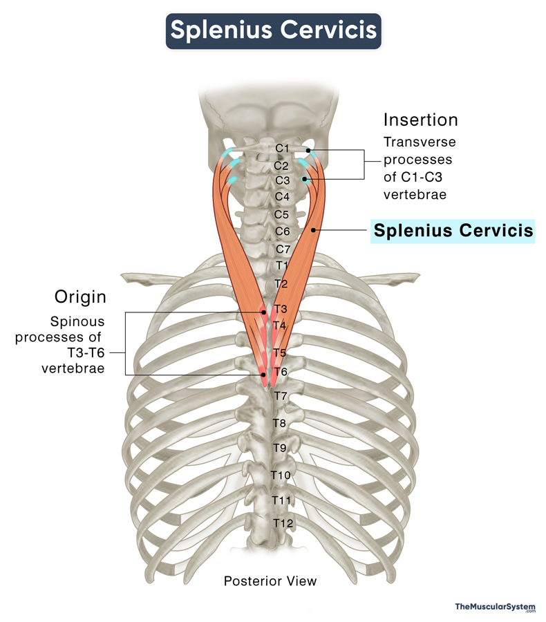
| Origin | Spinous processes of the T3 to T6 vertebrae |
| Insertion | Transverse processes of the C1 to C3 vertebrae |
Origin
The muscle originates in thin tendinous bands from the spinous processes of the 3rd to 6th thoracic vertebrae (T3 to T6).
Insertion
From the point of origin, the muscle fibers course obliquely upward and then curve inward toward the neck (cervical spine) to insert into the transverse processes of the 1st to 3rd cervical vertebrae (C1 to C3). Sometimes, part of the muscle may also insert into the 4th cervical vertebrae (C4).
Relations With Surrounding Muscles and Structures
The splenius cervicis, together with the splenius capitis, forms part of the superficial layer of the intrinsic back muscles. The cervicis muscle runs closely along the lower outer border of the capitis muscle, with the two almost blending at certain points, making them difficult to distinguish. Together, they overlie the deeper muscles in this group, lying immediately superficial to the capitis parts of the semispinalis and longissimus muscles.
Along with the other deep muscles in the neck region, splenius cervicis is wrapped by the investing layer of the deep cervical fascia, a connective tissue layer that surrounds and supports the structures in the neck.
The muscle also has the sternocleidomastoid (SCM) and the trapezius muscles lying superficial to it.
Function
| Action | Extending, rotating, and laterally flexing the head and neck |
The splenius cervicis muscle supports the neck and keeps it stable during movements. When the muscle contracts on one side (unilateral contraction), it helps rotate the neck or flex it if contracting on the same side. So, this is the muscle at work when you tilt your head or turn it to look over your shoulder.
When both sides of the muscle contract (bilateral contraction), it works with the trapezius muscle to straighten or extend the neck. It plays a vital role in extending and stabilizing the neck when standing up from a sitting position. This contraction helps position the neck so that the longus capitis, a muscle that primarily stabilizes the neck and cervical spine, can take over and bring your head back to a neutral, upright position once you are standing.
Antagonists
The sternocleidomastoid, the largest muscle at the front of the neck, can be considered antagonistic to the splenius cervicis as it is primarily responsible for bowing the head forward
and rotating it to the opposite side of the muscle’s contraction.
Innervation
| Nerve | Dorsal rami of the lower cervical nerves |
The muscle is innervated by the posterior or dorsal rami of the cervical spinal nerves, specifically those arising from the lower cervical vertebrae (usually from C3 and below).
Blood Supply
| Artery | Transverse and deep cervical arteries |
The muscle has an extensive blood supply coming from 5 different sources. These are:
- Transverse cervical artery
- Deep cervical artery
- Descending branch of the occipital artery
- Vertebral artery
- Superior intercostal artery
The venous drainage corresponds to the veins associated with these arteries.
References
- Splenius Cervicis Muscle: Radiopaedia.org
- Splenius Cervicis Muscle | Location, Action & Insertion: Study.com
- Splenius Cervicis: TeachMeAnatomy.info
- Splenius Cervicis: WheelessOnline.com
- Splenius Cervicis Muscle: Kenhub.com
- Splenius Cervicis Muscle: GetBodySmart.com
Della Barnes, an MS Anatomy graduate, blends medical research with accessible writing, simplifying complex anatomy for a better understanding and appreciation of human anatomy.
- Latest Posts by Della Barnes, MS Anatomy
-
Tensor Tympani
- -
Stapedius
- -
Auricularis Posterior
- All Posts


